3D Anatomy Model
Add another dimension to your learning with fully-interactive educational male and female anatomical models.
Learning about the human anatomy has never been more fun!
Purchase
The Back is a Musculoskeletal structure complex. It is made up of muscles, bones, nerves, and it also holds and protects different organs systems, for instance, the spinal cord. One of the most important supportive components of the back is the muscles present in it. These muscles play several roles e.g., movement of the torso, spine support, and coordination of arms and legs.
In the next paragraph, we will summarise the key information you need to know about the back muscles.
1. The back muscles are primarily divided into two groups: the extrinsic muscles and intrinsic muscles.
2. The extrinsic back muscles include the superficial back muscles which lie close to the skin. These extrinsic back muscles are responsible for the movement of the shoulder or upper limb. Thes muscles migrated to this region during fetal development and thus are also called migratory muscles. These muscles are further divided into superficial and intermediate groups.
3. The intrinsic back muscles, also called the true back muscle, are located beneath the extrinsic back muscles. The distinctive border between these two groups of muscles is a tough tissue known as the thoracolumbar fascia. The intrinsic back muscles are divided into superficial, deep, and deepest layers, and their main purpose is to produce movements.
4. All the muscles of the back are supplied by deep cervical, posterior intercostal, subcostal, or lumbar arteries.
5. The extrinsic muscles are innervated by the ventral (anterior) division of the spinal nerve whereas its posterior division innervates the intrinsic back muscles.
6. About muscle disorders: Back muscle pain can often be the result of a traumatic event, incorrect posture, or a nerve disorder.
Muscle imbalance can occur when the two symmetrical muscles on either side of the body have different sizes, strengths, or positioning.
Muscle strain, or pulled muscle, is often the result of a tearing or overstretching of a muscle due to an incorrect movement or its extensive use.
Disorders of the spine, the structures around the spine (joints, muscles, ligaments, nerve roots), or the disk between the vertebrae (i.e. herniated disc) can also cause back muscle pain.
7. Muscle relaxants (i.e. antispasmodics) are commonly used to treat muscle spasms, especially in chronic conditions such as fibromyalgia, a chronic neurological condition associated with general body pain.
Now, let’s deep dive into the structure and function of the back muscles!
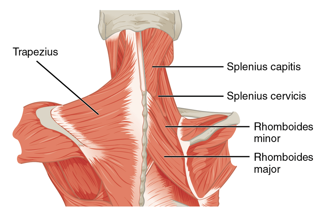
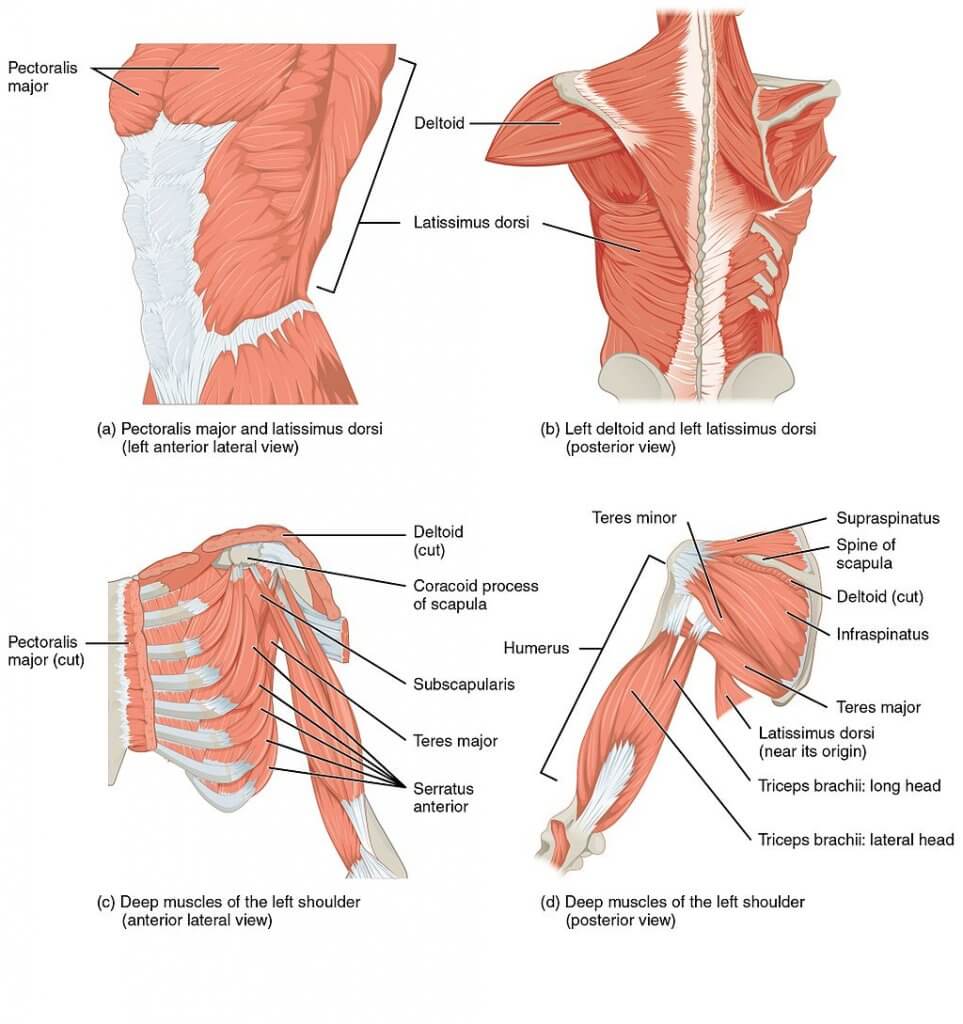
The extrinsic back muscles, as mentioned earlier, functionally belong to the upper limb muscles. They are:
The two major muscles in these groups are the trapezius (the traps) and latissimus dorsi. The traps are composed of three types of fibers: descending, ascending, and transverse fibers. They help us elevate, depress, or retract (pull back) the scapula.
The latissimus dorsi is the widest muscle of our body. The latissimus dorsi is the primary muscle that pulls you up on a pull-up bar.
The rest of the muscles have an additional supportive function and are displayed below in the diagram.
All the extrinsic back muscles are innervated by the ventral (anterior) division of the cervical spinal nerves. The trapezius muscle is an exception and it is supplied by the accessory nerve – the 11th Cranial Nerve (CN XI).
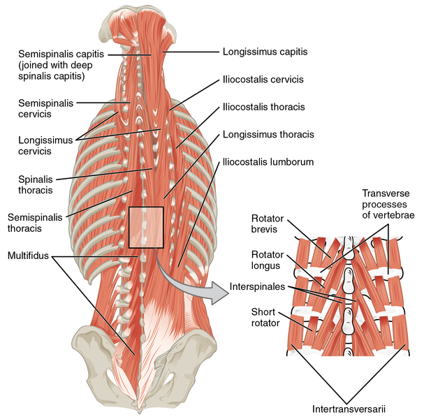
The intrinsic muscles of the back are the closest layer to the vertebral column. In the thoracic region, these muscles lie under the thoracolumbar fascia. While going below in the lumbar region, they are invested between the superficial and middle layer of the thoracolumbar fascia. These muscles specifically act on the vertebral column.
Intrinsic muscles of the back are divided into three groups:
The superficial layer includes the splenium and erector spinae muscles.
The erector spinae is a big muscle group composed of three muscular columns. It consists of the spinalis, longissimus, and the iliocostalis, and they are located bilaterally i.e., on either side of the spine.
The erector spinae muscles mostly attach between the transverse process of the vertebrae of the corresponding region. Each part plays a similar function and as a whole function to extends and laterally (sideways) flex the back. Extension is a bilateral action while lateral flexion function is a unilateral contraction acting on one side only.
Innervation – these muscles are innervated by the posterior division of the cervical spinal nerves.
The deep layer includes the following transversospinal Muscle: Semispinalis, Multifidous, and Rotatores.
The semispinalis muscle is further divided into semispinalis capitits, semispinalis cervicis, and the semispinalis thoracis (The above-motioned division is based on the location of these muscles around the spine).
These muscles contracting bilaterally (on both sides) extend the head, cervical and thoracic spine. They also produce lateral flexion of the head, cervical, thoracic spine and rotation of the head, cervical, and thoracic spine.
The multifidous muscle further consists of the multifidous cervicis, multifidous thoracis, and lumborum muscles. The multifidous group bilaterally acts to extend the spine, while unilateral contraction produces rotation and lateral flexion of the spine.
The rotatores muscles, finally, are divided into rotatores breves and longi. They act on the thoracic spine to extend and rotate it.
Innervation—Posterior division of the spinal nerves.
This is the deepest layer of back muscles and consists of two groups.
The Interspinales muscles are short muscles that extend between the spinous processes of the vertebrae. The function of this group is to contribute to the extension of the cervical and lumbar spine.
The Intertransversarii muscles attach to the transverse processes of the vertebrae. They are often absent in the thoracic region but are developed in the cervical and lumbar spine. Intertransversarii colli and intertransversarii lumborum are the two subdivisions of this group. These muscles contribute to the lateral flexion and stabilization of the spine.
Innervation – These muscles are supplied Posterior division of the spinal nerves
Muscles of the back are usually prone to physical trauma and other disorders. Lower back or neck pain is the most common complaint associated with the back muscles. It is commonly caused by inappropriate posture, long-duration sitting, and heavy-weight lifting. These disorders are mostly resolved by rest, physiotherapy, and the optional use of over-the-counter Nonsteroidal anti-inflammatory painkillers (NSAIDs).
The content shared on the Health Literacy Hub website is provided for informational purposes only and it is not intended to replace advice, diagnosis, or treatment offered by qualified medical professionals in your State or Country. Readers are encouraged to confirm the information provided with other sources and to seek the advice of a qualified medical practitioner with any question they may have regarding their health. The Health Literacy Hub is not liable for any direct or indirect consequence arising from the application of the material provided.


