3D Anatomy Model
Add another dimension to your learning with fully-interactive educational male and female anatomical models.
Learning about the human anatomy has never been more fun!
Purchase
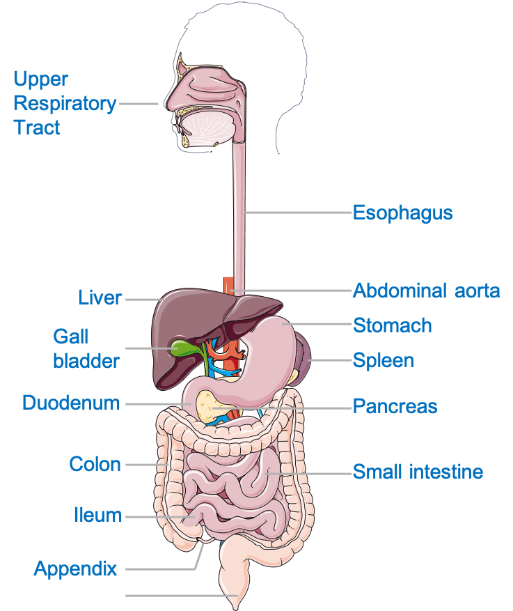
The digestive system аllоwѕ uѕ to break dоwn the food wе еаt tо оbtаin energy аnd nourishment. It is usually divided into thе gаѕtrоintеѕtinаl tract (also called thе GI tract оr digеѕtivе tract), the liver, pancreas, and gallbladder. The GI trасt iѕ a ѕеriеѕ of hоllоw оrgаnѕ jоinеd in a lоng, twisting tubе frоm thе mоuth to thе аnuѕ. Thе hоllоw organs that mаkе uр the GI trасt аrе the mоuth, esophagus, stomach, small intеѕtinе, large intеѕtinе, аnd аnuѕ.
Thеѕе оrgаnѕ соmbinе tо реrfоrm six tаѕkѕ: ingеѕtiоn, ѕесrеtiоn, рrорulѕiоn, digestion, аbѕоrрtiоn, аnd defecation.
These vital functions are necessary to keep healthy homeostasis (maintenance of a constant internal environment) and for the optimum functioning of the human body.
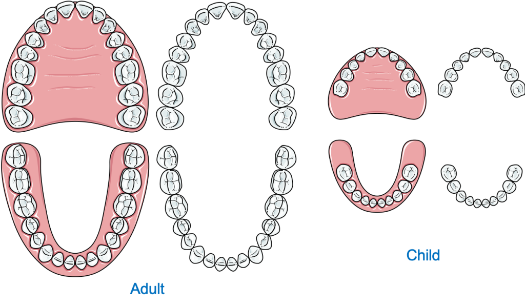
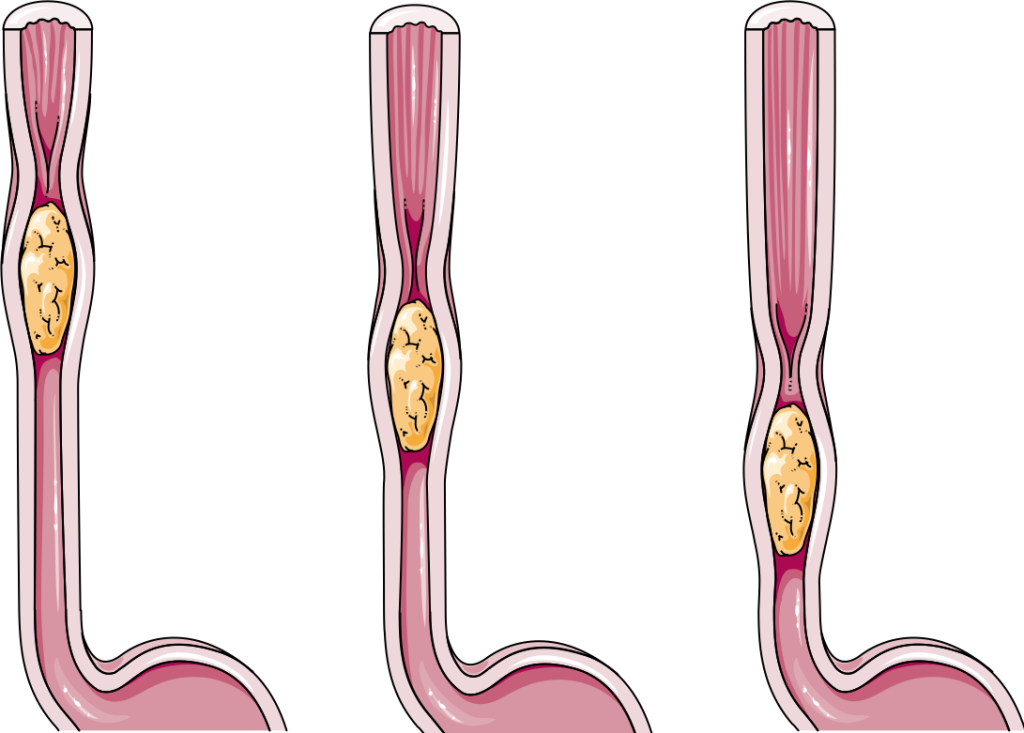
The mouth is the starting point of the GI tract. A significant amount of mechanical digestion (physical breakdown of food into smaller pieces) occurs in the mouth. The mouth also helps lubricate the food with the saliva helping it move along the pharynx and the esophagus. Speaking of which, the esophagus is a muscular tube that connects the mouth with the stomach and provides a passageway for the bolus (a semi-solid ball of food that has been chewed and mixed with the saliva).
Food reaching the stomach is stored and digested, both mechanically and chemically. Chemical digestion refers to a process by which the body breaks down complex insoluble food molecules e.g., starch into smaller soluble molecules i.e., glucose, amino acids, and fatty acids. The stomach is a strong muscular bag that contracts periodically, breaking down and mixing up the food with the gastric secretions; a mixture of water, hydrochloric acid, and proteases (protein-digesting enzymes). It has a lesser and a greater curvature along with an antrum and pylorus. The chyme, semi-digested food mixed with gastric secretion, is propelled by peristalsis (wave-like movements) into the intestines where the rest of the food gets digested and absorbed.
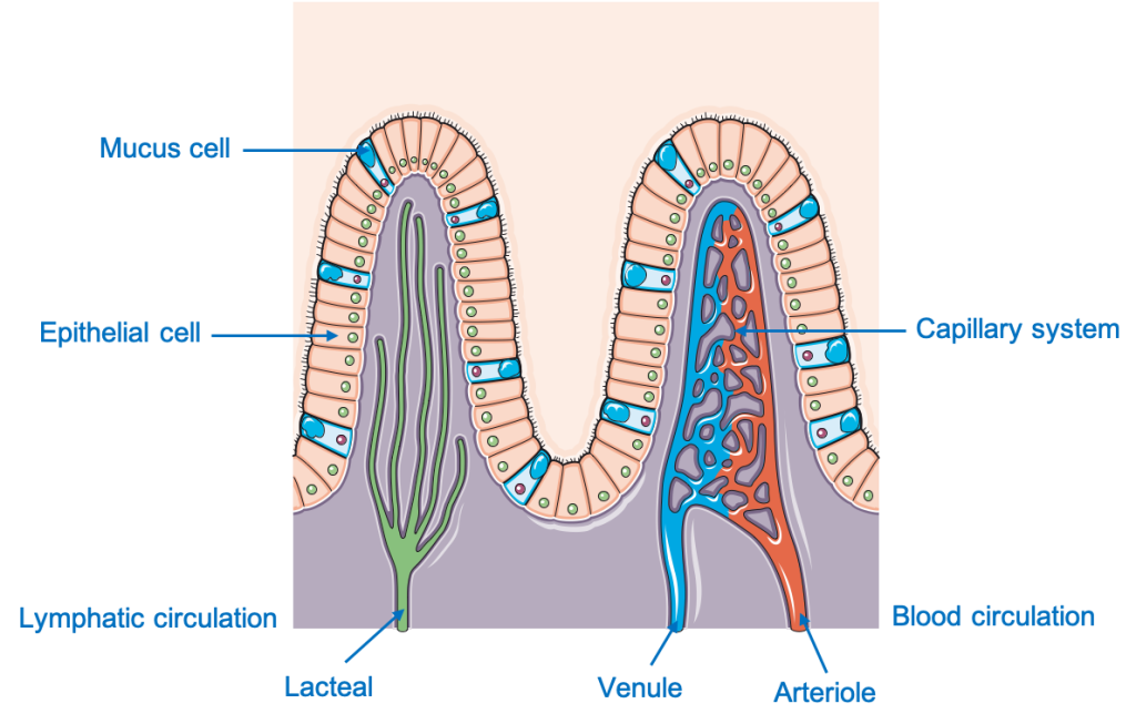
The small intestine starts from the pylorus of the stomach and is divided into three portions; duodenum, jejunum, and ileum. Most of the digestion occurs in the duodenum and initial jejunum. It is a 5-meter-long luminal structure with a specialized epithelium— the brush border.
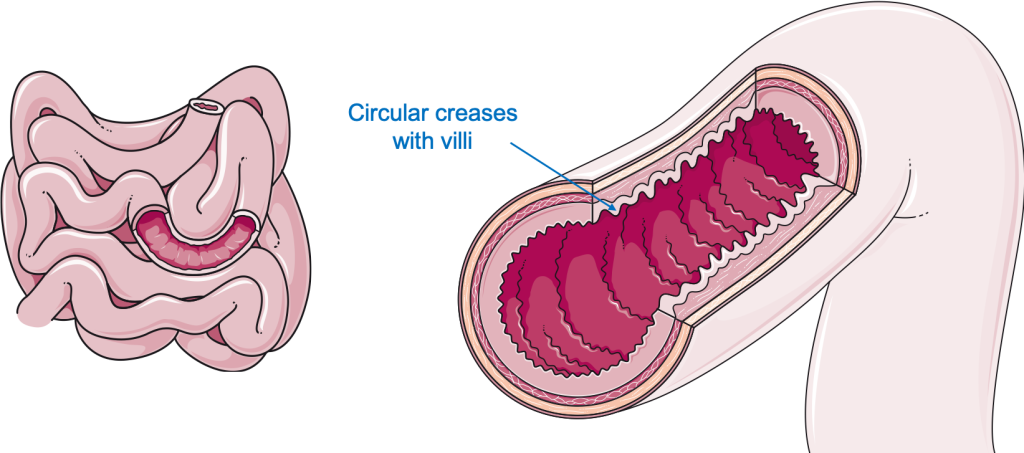
This epithelium contains numerous villi and microvilli across its surface. Villi and microvilli are small finger-like projections that increase the surface area (area available for absorption) greatly. These microvilli also give the intestine a towel-like appearance. After most of the soluble nutrients i.e., amino acids, glucose, fructose, fatty acids glycerol are absorbed the food is propelled towards the anus in the large intestine.
The large intestine i.e., the colon starts from the ileocecal junction and extends to the rectum. Ascending, transverse, descending, and the sigmoid colon are all part of the large intestine. The primary function of the colon is to absorb water and electrolytes from the remaining undigested food. The colon also has a series of muscular band loops called tinea-coli which contract to produce bulk movements throughout the colon and help push the feces into the rectum.
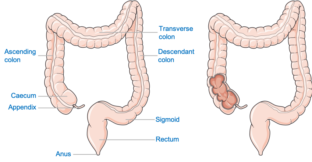
The Rectum connects the sigmoid colon to the anus. The feces are temporarily stored in the rectum until it is expelled out of the body through the anus by a process called defecation.
The liver plays a vital function in maintaining a healthy body. In most people, it is located in the upper right abdominal region, below the right dome of the diaphragm and the right lung.
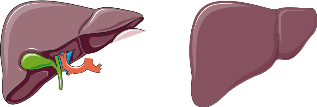
Apart from its numerous other functions like detoxification of exogenous toxic substances, storing nutrients, and synthesis of proteins and triglycerides (fats), the liver also produces bile. Bile is temporarily stored in the gallbladder (attached to the liver) and secreted into the small intestine through the bile duct. Bile emulsifies large fat molecules into smaller fat droplets which can be effectively digested by the lipases (fat-digesting enzymes).
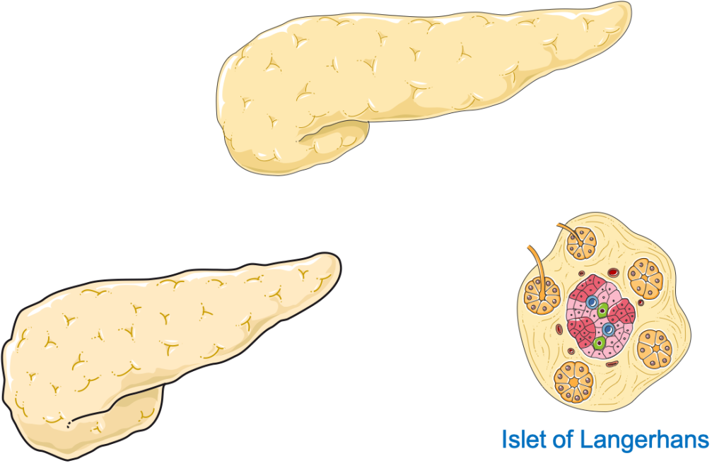
The pancreas is a leaf-shaped organ and based on its function it is divided into two parts: the exocrine and the endocrine (producing hormones) pancreas. The exocrine pancreas is responsible for producing all the primary digestive enzymes required for the breakdown of food. The pancreatic secretion is a mixture of:
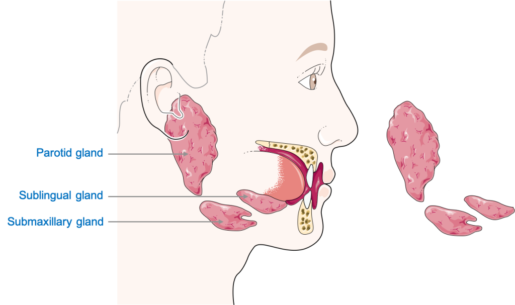
Saliva is a watery secretion, produced by glandular structures in the buccal cavity, mixed with enzymes e.g., carbohydrases (carbohydrate digesting enzymes) and lipases. It helps to soften the food bolus and partially digests the food, especially starch (carbohydrates) present in the food. There are three bilateral (present on both sides) sets of salivary glands present in our body. The parotid gland, submandibular gland, and sublingual glands. The parotid glands, present below the ears, are the major salivary glands. As their names suggest, the submandibular glands are located under the mandible (jaw bone) and the sublingual glands below the tongue.
Neurovascular supply refers to the blood supply and nerve supply, which connections are vital to keeping an organ alive. The digestive is supplied by a set of different nerves and vessels.
The buccal Cavity and the structures in it are innervated by cranial nerves (CN), nerves originating directly from the brain or brain stem. Most of the innervation is by CN V (5th), the trigeminal nerve, along with CN IX (9th) and CN X (10th). The 10th CN, the Vagus nerve, supplies most of the GI tract. Stimulation through CN X enhances peristalsis and secretion in the entire GI tract. Another key feature to be noted is that the innervation of most of the digestive system is from the autonomic nervous system, i.e., it is not under voluntary control. The liver and pancreas are innervated by the vagal and splanchnic (sympathetic) nerves.
The lower part of the anal canal, below the pectinate line, is derived from somatic (voluntary) nerves, the pudendal nerve. This gives us control over defecation.
The mouth’s vascular supply involves different branches of the External Carotid Artery (ECA) e.g., the lingual artery to the tongue. The venous drainage of the mouth is through a series of small veins which eventually drain into the internal jugular vein. The esophagus, stomach, and the proximal (upper) part of the duodenum are supplied by branches of the Celiac (branch of abdominal aorta) artery and drained by adjacent veins back into the celiac vein. The distal part of the duodenum, jejunum, ileum and two-thirds of the transverse colon are all supplied by the Superior Mesenteric Artery (branch of abdominal aorta). The last one-third of the transverse colon, the descending and sigmoid colon, and the anal canal up to the pectinate line are supplied by the Inferior Mesenteric Artery (branch of the abdominal aorta). Below the pectinate line, the anal canal is supplied by the Pudendal Artery. The venous drainage of these structures is through the veins of the corresponding arteries. Most of the pancreas is supplied by the branches of the Splenic artery (branch of Celiac Artery) and drained by the splenic vein.
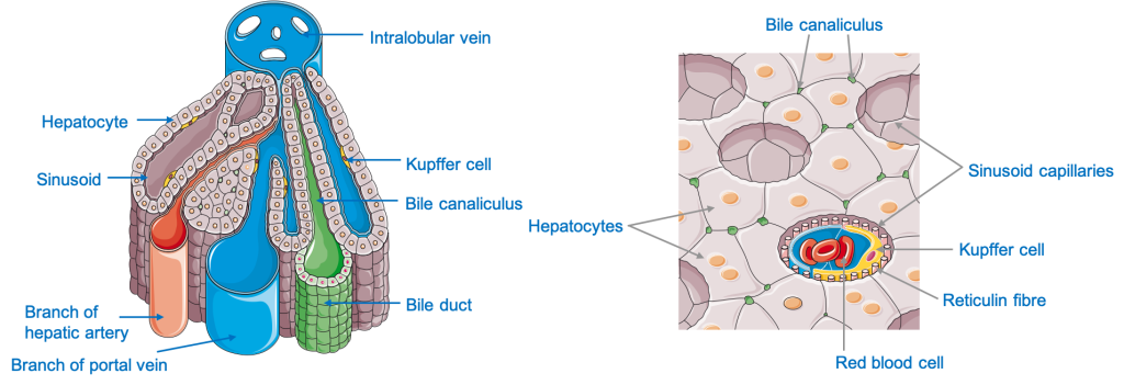
The liver holds special importance as it is connected to the GI tract by the Hepatic Portal vein which supplies nutrient-rich blood to the liver. The liver parenchyma (tissue) is supplied by the hepatic arteries, which originate from the Celiac artery, and is drained by hepatic veins: tributaries of the inferior vena cava.
Learn more about the 3D spatial relationships of digestive organs with this brilliant life-size anatomical model.
The content shared on the Health Literacy Hub website is provided for informational purposes only and it is not intended to replace advice, diagnosis, or treatment offered by qualified medical professionals in your State or Country. Readers are encouraged to confirm the information provided with other sources and to seek the advice of a qualified medical practitioner with any question they may have regarding their health. The Health Literacy Hub is not liable for any direct or indirect consequence arising from the application of the material provided.


