3D Anatomy Model
Add another dimension to your learning with fully-interactive educational male and female anatomical models.
Learning about the human anatomy has never been more fun!
Purchase
The mediastinum is a critical anatomical region of the human body. However, this is not an organ. Therefore, let’s start by summarizing the five key facts about the mediastinum before deep-diving into the details of this body part.
1. The mediastinum is not an organ but is a compartment in the thorax that hosts a number of organs, vessels, nerves, and other structures.
2. The mediastinum hosts the most vital organs of the human body: the heart.
3. The mediastinum is virtually divided into anterior, middle, posterior, and superior regions. This division is mainly used for clinical purposes.
4. The mediastinum is one of those regions in the body that has different borders: anterior (defined by the manubrium of the sternum), posterior (defined by the thoracic vertebrae), superior (defined by the superior thoracic aperture or outlet), inferior (defined by the diaphragm), and lateral (defined by the lungs and each pleural cavity).
5. The anatomical division of the mediastinum has been largely debated in the past. While radiologists used to define this space based on the landmarks observed in the radiological images, surgeons were relying on the intraoperative margins. A CT scan-based division is now the most common and consistent approach across different medical specialties.
6. The primary function of the mediastinum is to provide these structures with a protected pathway.
7. The mediastinum is also clinically significant due to many pathologies occurring in it. One of the most invasive and difficult to treat is a mediastinal tumor. A mediastinal mass can be found in the anterior, posterior, or middle mediastinum, and depending on their location they can apply pressure on vital organs such as the lungs, esophagus, spine, or trachea, leading to serious consequences. If a mediastinal tumor is suspected, the practitioner will recommend different tests, including a biopsy of the mediastinal mass in the thoracic cavity, a ct scan, or other imaging procedures and functional tests. Depending on the size and positioning of the tumor, surgical resection can be an option and it can be combined with pharmacological intervention to reduce the size of the mass and limit the risk of metastasis.
Let’s now explore the anatomy and function of the mediastinum in the sections below.
Explore the mediastinum and its organs in 3D, AR, and VR with the interactive model below.
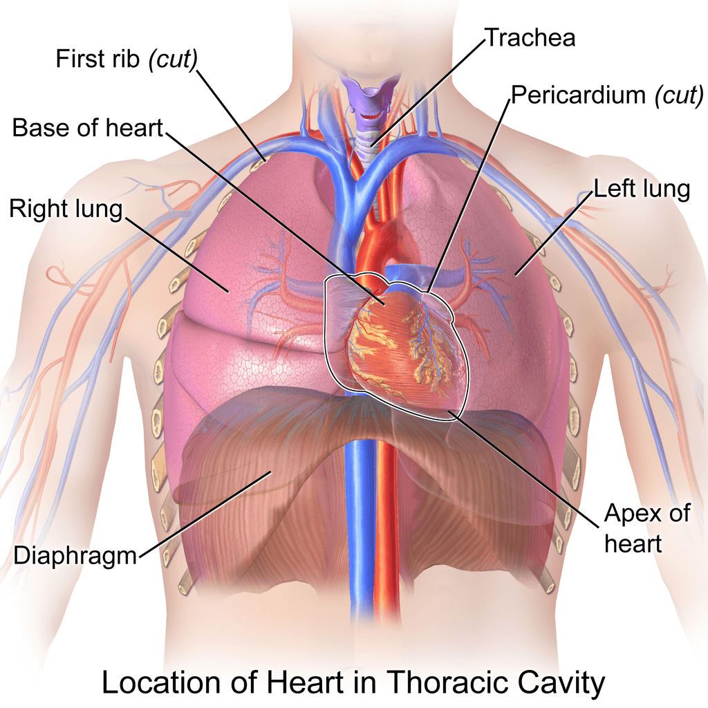
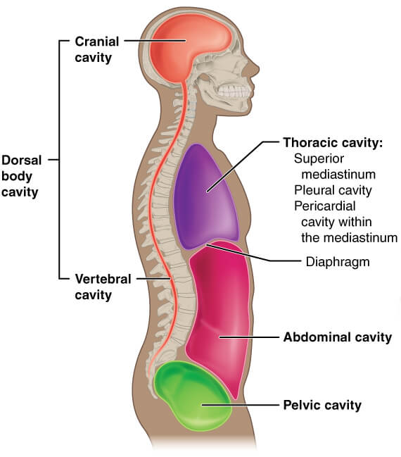
The mediastinum is the space between the two lungs that extends the whole length of the thoracic cavity. From the top to the lowest point, the mediastinum extends from the thoracic inlet to the upper lining of the diaphragm. Though there is no distinct physical partition of the mediastinum, it is divided into the superior and inferior.
The inferior mediastinum is further divided into anterior, middle, and posterior compartments. Hence, there is a total of four compartments of the mediastinum. The Mediastinum houses vital body structures, such as the heart, esophagus, trachea great vessels, and several nerves.
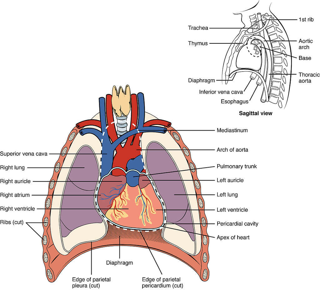
The uppermost margin of the superior mediastinum is the thoracic inlet, and the inferior boundary is the thoracic plane or sternal angle. The pleural sacs bound the superior mediastinum laterally. The anterior border is the dorsal surface of the sternum, while the posterior boundary is the central aspect of the first four thoracic vertebrae.
Superior mediastinum houses organs, blood vessels, and nerves. The organs of the superior mediastinum include the thymus, esophagus, and trachea. The blood vessels of the superior mediastinum include the aortic arch, brachiocephalic trunk, and left subclavian and left carotid artery. The veins of the superior mediastinum include SVC, brachiocephalic vein, and azygous vein. It also contains some parts of the thoracic lymphatic duct. The nervous content o the superior mediastinum consists of both vagus nerves, phrenic nerves, recurrent laryngeal nerves, and cardiac nerves.
The inferior mediastinum extends from the lower boundary of the superior mediastinum to the upper surface of the diaphragm. It is divided into anterior, middle, and posterior compartments.
The contents of the anterior mediastinum include the thymus, branches of internal thoracic vessels, and parasternal lymph nodes.
The middle mediastinum contains the heart, major blood vessels, i.e., aorta, pulmonary trunk, SVC, and pulmonary veins. A part of the vagus, phrenic, and sympathetic nerves also passes through the middle mediastinum.
The contents of the posterior mediastinum include the esophagus and descending thoracic aorta. It also contains part of the thoracic duct, azygous and hemizygous veins. It also has part of the vagus, splanchnic, and sympathetic chain nerves.
The primary function of the mediastinum is to provide a safe passage to different structures through the thoracic cavity and house the essential organs of the thoracic cavity.
The main function of the superior mediastinum is to create a conduit or passage for different structures from the head & neck to the thoracic cavity. The main function of the anterior mediastinum is to protect the structures lying behind it. It has connective tissue and fat that acts as a cushion against the external thrust.
The middle mediastinum hides the heart and great blood vessels behind the shield of the Anterior Mediastinum. The posterior mediastinum is considered a continuation of the superior mediastinum. The posterior mediastinum is a conduit for the structures traversing from the thoracic cavity to the abdominal cavity.
The blood supply, venous drainage, and Nerve supply of the different mediastinum compartments are from the neurovascular content of the respective compartment. The visceral organs of the mediastinum are also supplied by the nerves and vessels of their respective compartments.
The mediastinum is the vital space of the body in the clinical aspect. It has several cardiopulmonary structures. The pathologies and abnormalities of the visceral contents of the mediastinum correlate with different compartments of the mediastinum. A brief description of the clinical importance of mediastinum is in the following section.
The mediastinum is the vital space of the body in the clinical aspect. It has several cardiopulmonary structures. The pathologies and abnormalities of the visceral contents of the mediastinum correlate with different compartments of the mediastinum. A brief description of the clinical importance of mediastinum is in the following section.
The superior mediastinum acts as a conduit for many vessels and nerves. The penetrating wounds of the upper part of the chest are likely to damage the vessels and nerves of the superior mediastinum. The upper mediastinum contains the arch of the aorta, which is a potential site of an aortic aneurysm. If an aneurysm of the aortic arch goes undiagnosed, it can lead to death.
The superior mediastinum also contains a part of the esophagus and trachea. The obstruction of the esophagus and trachea leads to severe results. The obstruction of the trachea causes respiratory compromise and subsequent death.
The main clinical importance of the anterior mediastinum is related to the disease of the thyroid gland, which nests in its upper part. The anterior mediastinum is also susceptible to crush injuries of the chest. These crush injuries may result in fat necrosis and hemorrhage in the anterior mediastinum.
The middle mediastinum is the clinically most important compartment of the mediastinum. It contains the heart and roots of great blood vessels. The heart diseases such as myocardial infarction, cardiac tamponade, and cardiomyopathy are related to the middle mediastinum. The congenital abnormal of the heart and great vessels are also associated with the middle mediastinum.
The posterior mediastinum is an ascending route for a lot of structures. The pathologies of the middle mediastinum can cause systemic dysfunction. Aneurysm of descending thoracic aorta, obstruction of the thoracic duct, and esophageal dysplasia are notorious clinical conditions related to the posterior mediastinum that readily evolve into life-threatening conditions.
The posterior mediastinum is an ascending route for a lot of structures. The pathologies The mediastinum is the space or cavity that extends between the pleura of two lungs. From superior to inferior, the mediastinum extends from the thoracic inlet to the superior surface of the diaphragm. The mediastinum extends between the sternum’s dorsal surface and the vertebral column’s ventral surface from the anterior to the posterior. The mediastinum is arbitrarily divided into four components; superior, anterior, middle, and posterior compartments. Each compartment contains different visceral organs, blood vessels, nerves, and lymphatics. The neurovascular of the mediastinum is from its content blood vessels and nerves. The mediastinum is clinically significant due to the visceral structure and blood vessels it contains. The middle mediastinum is clinically most important due to the presence of the heart in it.
1. Stoddard N, Heil JR, Lowery DR. Anatomy, Thorax, Mediastinum. [Updated 2020 Jul 31]. In: StatPearls [Internet]. Treasure Island (FL): StatPearls Publishing; 2021 Jan-. Available from: https://www.ncbi.nlm.nih.gov/books/NBK539819/
2. Priola, S. M., Priola, A. M., Cardinale, L., Perotto, F., & Fava, C. (2006). The anterior mediastinum: anatomy and imaging procedures. La Radiologia medica, 111(3), 295–311. https://doi.org/10.1007/s11547-006-0031-6
3. Rizvi, S., Wehrle, C. J., & Law, M. A. (2020). Anatomy, Thorax, Mediastinum Superior and Great Vessels. In StatPearls. StatPearls Publishing. https://pubmed.ncbi.nlm.nih.gov/30137860/
4. Stoddard, N., Heil, J. R., & Lowery, D. R. (2020). Anatomy, Thorax, Mediastinum. In StatPearls. StatPearls Publishing. https://pubmed.ncbi.nlm.nih.gov/30969641/
5. Mast, W. R., & Jafek, B. W. (1975). Mediastinal anatomy for the mediastinoscopist. Archives of otolaryngology (Chicago, Ill. : 1960), 101(10), 596–599. https://doi.org/10.1001/archotol.1975.00780390010003
The content shared in the Health Literacy Hub website is provided for informational purposes only and it is not intended to replace advice, diagnosis, or treatment offered by qualified medical professionals in your State or Country. Readers are encouraged to confirm the information provided with other sources, and to seek the advice of a qualified medical practitioner with any question they may have regarding their health. The Health Literacy Hub is not liable for any direct or indirect consequence arising from the application of the material provided.


