3D Anatomy Model
Add another dimension to your learning with fully-interactive educational male and female anatomical models.
Learning about the human anatomy has never been more fun!
Purchase
The human brain is a very sensitive structure in terms of trauma and injury. Several protective features help keep the brain in a protective environment. One of these features is the meninges.
Let’s have a look at the most interesting facts about the meninges.
1. The meninges are sheet-like covering of the brain that help keep the Central Nervous System ( CNS ) in place and stabilise it. The meninges are also found within the spinal canal in the spinal cord.
2. In the brain and spine, the meninges also regulate the circulation of cerebrospinal fluid (CSF) that provides protection and nutrients to the nervous tissue.
3. Specialised cells (meningeal cells) in the meningeal tissue are responsible for a protective mechanism called meningeal immunity that controls and intervene in the presence of a pathogen-mediated infection.
4. What we refer to as “the meninges” represents three independent layers of tissue located in the skull (cranial cavity) and surrounding the spinal cord characterised by a unique structure and role. The dura mater, the outermost layer, consists of two sheets of connective tissue lining the interior surface of the cranial cavity. The arachnoid layer, the middle layer, is made of non-vascularised connective tissue. The pia mater, the innermost layer, is a very thin sheet that covers the brain surface and is highly vascularised.
5. The space between the arachnoid and pia maters is known as “subarachnoid space”. In this region where the cerebrospinal fluid (CSF) circulates, there is a particular compartment (subarachnoid cisterns) characterised by the accumulation of CSF (pools or cisterns) to provide the brain with nutrients, enable solute exchange, and offer mechanical and functional support to the brain.
In this article, we will discuss in detail the structure, function and most common diseases associated with this brain-protective layer.
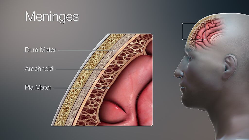
Our brain is suspended or floating in the CSF and covered by three layers of the meninges:
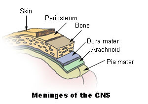
The meninges envelop both the brain and the spinal cord and separate them from the bones surrounding them i.e., the skull and the vertebral column. The meninges are further topographically classified into cranial and spinal meninges. There are three clinically important spaces bounded by the meninges, they are the epidural, subdural, and subarachnoid spaces. The arachnoid and pia mater are also called leptomeninges; the reason being the presence of CSF between these two layers.
Apart from its mechanical function, the meninges act to support the blood vessels and form a continuous cavity for the circulation of cerebrospinal fluid.
The dura mater is the outermost meningeal layer. It consists of dense irregular connective tissue and it is composed of two layers:
These two layers are inseparable except in places where they separate to envelop large veins known as dural venous sinuses. In these above-mentioned places, the dura mater projects inward towards the brain tissue forming a fibrous partition. These partitions are within the cranium and are as follows:
The spinal part of the dura mater contrastingly does not have the periosteal layer. This difference can be attributed to the fact that unlike the skull the vertebral column has its own periosteal layer.
This layer of meninges is sandwiched between the dura and pia mater. The potential space between the dura and arachnoid mater is named the subdural (deeper to dura) and the space between the arachnoid and pia mater is called the subarachnoid (deeper to arachnoid) space. The latter is a true space that contains the Cerebrospinal fluid in it. This space is also the location of all the cerebral arteries and veins. The spinal arachnoid matter is the continuation of the cranial arachnoid mater.
The arachnoid is attached to the dura and pia mater at different places. At the site of attachment with the dura matter, there are mushroom-like protrusions called the arachnoid granulations. These granulations protrude into the dural venous sinuses and enable the continuous flow of CSF from the subarachnoid space to the venous system.
It is a thin, highly vascular layer that closely follows the brain contours. Some vessels of the brain are related to this layer but they are embedded in the pia mater and are not named separately. The pia mater functions to physically separate the nervous tissue from the vasculature present in the subarachnoid space. It also increases the efficacy of the blood-brain barrier, a vital component for the brain tissue to stay healthy.
The spinal pia mater closely attaches to the spinal cord and gives off a fibrous projection at the end of the spinal cord – the filum terminal.
As described earlier the three meningeal layers have potential spaces between them and are as follows:
These spaces are of much clinical importance in traumatic injuries to the head. Blood can leak into these spaces pathologically and cause haemorrhages which can be fatal.
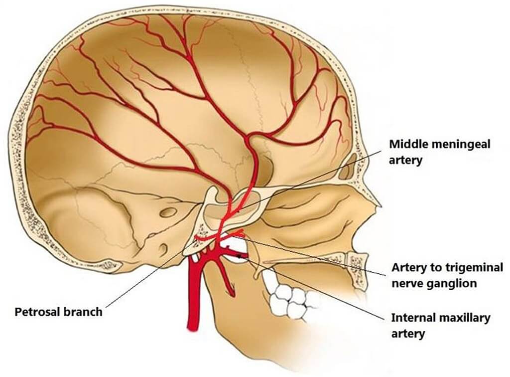
The meninges are innervated by the Trigeminal Nerve (Fifth cranial nerve). The blood supply to the meninges is from the branches of the carotid and vertebral arteries.
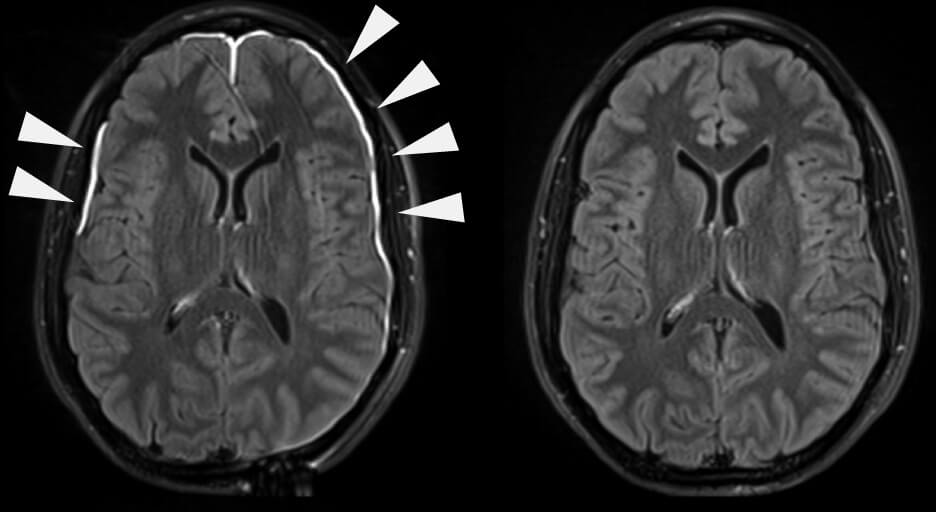
The inflammation (swelling) of the meninges is termed meningitis. It is most commonly caused by bacteria but can be caused by viruses and is even drug-induced. Bacterial meningitis is most commonly caused by two bugs (pathogens): Neisseria meningitidis and Streptococcus pneumonia. Viral meningitis, on the other hand, is most commonly caused by the enteroviruses.
Meningitis is usually treated by antibiotics and antiviral medication after confirming the cause. In untreated, however, can progress towards serious complications e.g., brain herniations, etc.
Learn more about meningitis in this article.
A haematoma is blood pooling or collection. Increased pressure in the skull due to this build-up causes a rapid increase in the intracranial pressure due to the skull being a closed cavity. On CT scans they are seen as crescent-shaped formations. There are two types of haematomas:
Another classification of subdural haematomas is based on the timeframe of their formation i.e., acute or chronic subdural haematomas. Acute haematomas are caused by sudden forceful brain injuries while chronic haematomas can result from a weak or small injury and are usually due to another underlying cause. Haematomas can be fatal if they are not treated immediately.
The content shared in the Health Literacy Hub website is provided for informational purposes only and it is not intended to replace advice, diagnosis, or treatment offered by qualified medical professionals in your State or Country. Readers are encouraged to confirm the information provided with other sources, and to seek the advice of a qualified medical practitioner with any question they may have regarding their health. The Health Literacy Hub is not liable for any direct or indirect consequence arising from the application of the material provided.


![SEER Development Team[1], Public domain, via Wikimedia Commons](http://training.seer.cancer.gov/module_anatomy/images/illu_meninges.jpg)