3D Anatomy Model
Add another dimension to your learning with fully-interactive educational male and female anatomical models.
Learning about the human anatomy has never been more fun!
Purchase
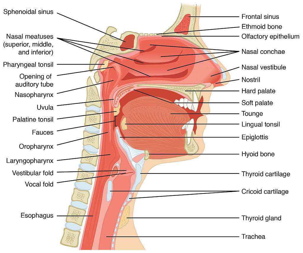
The nasal cavity is a large, air-filled space that opens at the nostrils, which are the two openings of the nose. The nasal septum is a partition that divides the cavity into two separate cavities. The nasal cavity is lined with a mucus membrane that helps keep your nose moist. The openings to the nasal cavity at the nostrils are lined by modified skin and have short hairs called vibrissae. The nasal cavity has many important anatomical landmarks. It has a roof (the uppermost part of the cavity), projections of mucosa from either side called concha, and small empty spaces (sphenoethmoidal recess) which are lined with olfactory mucosa. These barriers give mechanical protection from the invasion of pathogens.
In this article, we will cover everything you need to know about the nasal cavity and what are the most common diseases associated with this body part.
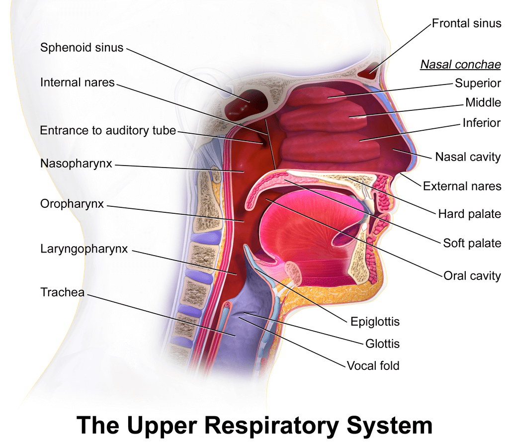
This cavity is divided into two separate cavities by the nasal septum. Each cavity consists of a roof, floor, and two walls on the sides; medial wall, and lateral wall. Each cavity comprises a nasal vestibule (which is the area below the nostrils), respiratory section, olfactory(smell-related) region, and some surrounding structures.
The area just below the nostrils is called vestibule, lined with course hairs which filter dust, sand and other particles keeping them from entering the lungs.
This section of the cavity refers to the passages through which air travels to reach the trachea and eventually the lungs. The respiratory section of each nostril contains four conchae (protrusions) covered by the nasal mucosa. Underlying these conchae are small holes. These openings connect to the paranasal sinuses surrounding the cavity.
The olfactory mucosa is located in the upper region of the nasal cavity, supporting the olfactory mucus membrane for the sensation of smell.
The surrounding structures include:
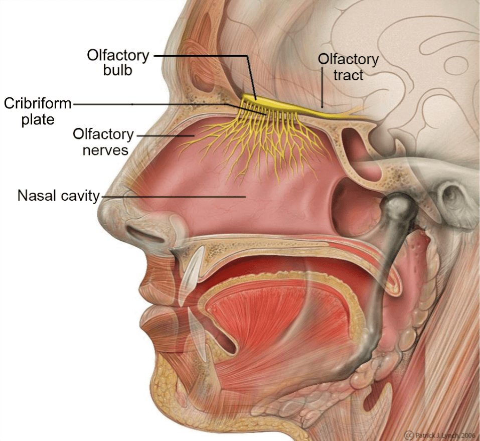
Olfaction is the sense of smell that happens at the apex of the cavity (olfactory area). This portion of the nasal cavity is lined with the cells called exteroception epithelial tissue, and it is interspersed with neurons containing sensory cilia.
The pathway of olfactory conduction goes from the olfactory receptor cells to the olfactory bulb in the forebrain, via the olfactory nerve.
Sensations of smell are experienced when certain chemical substances become dissolved in the thin layer of fluid covering the surface of the mucous membrane of special cells called olfactory receptor cells which come in contact with the olfactory hairs, or cilia.
The inhaled air must be warmed and humidified before it reaches the lungs. This is mainly done in the upper respiratory portion of the nasal cavity which is lined with special cells known as the ciliated pseudostratified epithelium.
The moisture of the mucus plays a role in the humidification of inhaled air. Also, the turbinates slow down the airflow keeping the air in the nasal passageways long enough to be warmed and humidified.
The cilia in the nasal cavity also play a role in defending the organism, as they move the mucus and eventual pathogens (germs and bacteria) towards the throat (pharynx) to eliminate them.
The nose receives blood from the internal and external carotid artery arteries.
The internal carotid artery branches to supply:
• Anterior ethmoidal artery
• Posterior ethmoidal artery
The ethmoidal arteries descend into the cavity through openings in the frontal bone (the cribriform plate.) to reach the nasal cavity.
External carotid branches that supply the nasal cavity include:
• Sphenopalatine artery
• Greater palatine artery
• Superior labial artery
• Lateral nasal arteries
The innervation of the nose is often functionally divided into special and general innervation.
Unique sensory innervation refers to the power of the nose to smell. The exteroception nerves are responsible for this. Branches of the first cranial nerve run through the cribriform plate to produce special sensory innervation to the nose.
General sensory innervation to the septum and the lateral walls is delivered by the nasopalatine nerve and the nasociliary nerve (branch of the ophthalmic division of the trigeminal nerve; the fifth cranial nerve.)
The inflammation and swelling of the mucus membrane of the nose. It is characterized by a runny nose, sneezing, and stuffiness.
It can be treated by:
Epistaxis is simply an elaborated medical term for a bloody nose. The nasal cavity is very vascularized so any trauma to the nose, dry nasal passageways, certain medications like blood thinners, or chronic conditions like a sex-linked disorder can commonly cause bleeding from a part of the nasal septum called the Little’s area.
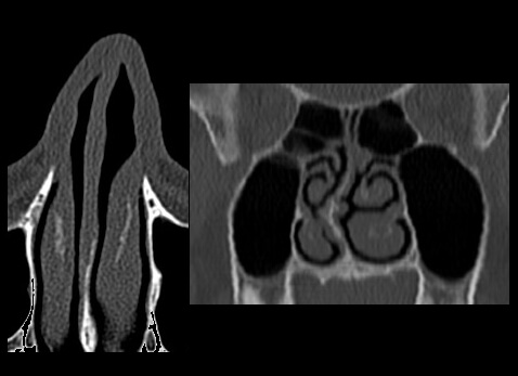
A deviated septum occurs when the thin wall (nasal septum) between your nasal passages is displaced to one side. A deviated septum can develop in utero, or while the fetus is still in the womb, as well as during the birthing process.
The presence of a deviated septum can cause frontal headaches and bleeding from the nose.
Turbinates are small structures inside the nose that cleanse and humidify the air that passes through the nostrils into the lungs. They are made of bone and soft tissue and are located inside the nose near the septum. Enlarged turbinates are a reaction to seasonal allergens. Sometimes, the enlargement is caused by environmental irritants. Chronic sinusitis, which causes persistent inflammation in the nasal passages, may also trigger chronic swelling of the turbinate. The treatment for enlarged Turbinates is often surgical reduction.
Marie nut. Essentials of Human Anatomy and Physiology. (7th edition). San Francisco: Benjamin Cummings; 2003. [Book]
Kaiser GE. The innate system. Doc Kaiser’s biology Home Page. CommunityCollegeBaltimore County. 2007.
Orahilly R, Muller F, Carpenter S, Swenson R. The nose and bodily cavity sinuses. Chapter 52. Basic Human Anatomy. R Swenson, Ed. DartmouthMedical college. 2008.
Mann MD. sense impression and exteroception senses. Chapter 10. The system in action. [online]. 2008. University of American state Medical Centre. [Book]
Oneal RM, Beil Jr RJ, Schlesinger J. Surgical anatomy of the nose. Otolaryngol Clin North Am. 1999 Feb;32(1):145-81. [PubMed]
Patel RG. Nasal Anatomy and performance. Facial Plast Surg. 2017 Feb;33(1):3-8. [PubMed]
Lafci Fahrioglu S, VanKampen N, Andaloro C. StatPearls [Internet]. StatPearls Publishing; Treasure Island (FL): Gregorian calendar month eleven, 2020. Anatomy, Head and Neck, Sinus perform and Development. [PubMed]
The content shared in the Health Literacy Hub website is provided for informational purposes only and it is not intended to replace advice, diagnosis, or treatment offered by qualified medical professionals in your State or Country. Readers are encouraged to confirm the information provided with other sources, and to seek the advice of a qualified medical practitioner with any question they may have regarding their health. The Health Literacy Hub is not liable for any direct or indirect consequence arising from the application of the material provided.


