3D Anatomy Model
Add another dimension to your learning with fully-interactive educational male and female anatomical models.
Learning about the human anatomy has never been more fun!
Purchase
The nose is the respiratory and olfactory organ of the body. The nasal cavity consists of several compartments and structures such as conchae, meatuses, cribriform plates, and many more.
Nasal turbinates or nasal conchae are the narrow, long, and curled bones which protrude inside the nasal cavity. These bony projections run towards the midline and downwards in the nasal cavity. All types of conchae help in dividing the nasal cavity into groove-like air passages that are essential for nasal breathing. In addition, these bones help provide significant landmarks for the surgical procedures that involve accessing the skull through the nose.
Nasal conchae divide into three parts:
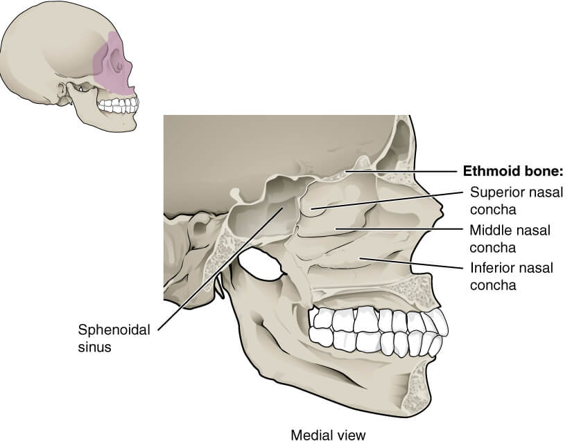
Meatuses are the passages that lie underneath each conchae. Each of these meatuses is capable of communicating with the nasal cavity independently
Meatuses are divided into 3 types:
It lies under the surface of the inferior concha, and of all the three meatuses, the inferior concha is the largest. A duct that drains tears from the eyes, known as the Nasolacrimal duct, connects to the inferior meatus and drains into the nasal cavity.
It lies just below the middle concha. The middle meatus is connected to multiple air sinuses on the face.
It lies just below the superior concha. Of all the meatuses, superior meatus is the shallowest and shortest meatus in size. This is because the superior meatus receives openings from the ethmoidal sinuses.
The four quadrants of the nasal conchae are supplied by sensory nerve branches of the trigeminal nerve
Olfactory or special sensory nerves have distributions to the superior concha to aid in the sense of smell.
The nasal cavity and surrounding structures have a rich supply of blood. Both the external and internal carotid arteries give blood supply to the nose.
Ethmoidal arteries are the ophthalmic artery branches that descend into the cavity of the nose via the cribriform plate to supply the nasal concha.
The facial artery also gives blood supply to parts of the conchae. A few branches of the sphenopalatine artery also supply the concha.
The nasal conchae form a venous plexus that drain to the facial, and pharyngeal veins.
For lymphatic drainage, lymph from the nasal conchae drains into the submandibular, deep cervical, and retropharyngeal nodes.
There are a few basic functions of the nasal conchae:
Invasion of the nasal cavity or concha by microorganisms causes infection of the nasal cavity This can be acute, chronic, or recurrent acute depending on the duration. Infection can lead to abscess formation. Most nasal cavity infections are viral, and only a small proportion develops due to bacteria. Rhinoviruses, influenza viruses, and parainfluenza viruses are the most common causes of sinusitis (inflammation of the sinuses).
Acute invasive fungal sinusitis is the infection of sinuses by fungi. This type of infection is common in people having immunocompromising conditions like HIV, Organ transplant patients, and Diabetes. The responsible pathogens for this infection are Absidia, Rhizopus, and Aspergillus.
JNA is a vascular mass commonly found in the nasal cavity of adolescent males. These growths are not cancerous but can damage nerves and bones in the nasal cavity. They can also block the ear and sinus drainage. The commonest causes of the JNA are nasal obstruction and recurrent nasal bleeding. The mainstay of treatment for juvenile nasal angiofibroma is surgery to remove the tumor.
This is the inflammation of the nasal cavity structures. It causes nasal blockage, frequent sneezing, and excessive water-like discharge from the nose. These symptoms happen when the person breathes in something they are allergic to, such as dust, animal dander, or pollen.
The most common characteristic of this condition is the hypertrophy (increase in size) of the inferior conchae causing nasal obstruction and difficulty in breathing.
Fractures of the nasal bone are commonly due to physical or mechanical trauma. Fracture of the nasal bone can lead to profuse bleeding and obstruction in the nasal cavity. Fractures associated with the nasal cavity are treated by Ear, Nose and Throat surgeons.
The content shared on the Health Literacy Hub website is provided for informational purposes only and it is not intended to replace advice, diagnosis, or treatment offered by qualified medical professionals in your State or Country. Readers are encouraged to confirm the information provided with other sources and to seek the advice of a qualified medical practitioner with any question they may have regarding their health. The Health Literacy Hub is not liable for any direct or indirect consequence arising from the application of the material provided.


