3D Anatomy Model
Add another dimension to your learning with fully-interactive educational male and female anatomical models.
Learning about the human anatomy has never been more fun!
Purchase
The neck is the structure that connects your head with your trunk and is essential for performing routine activities. The neck acts as a point that provides mobility and flexibility to your head and it can be moved in different directions.
The neck is divided into four main compartments; vertebral, visceral, and a pair of vascular compartments. These compartments of the neck contain cervical vertebrae, the cervical part of the spinal cord, parts of the digestive and respiratory system, blood vessels, and nerves. Muscles of the neck are not included in the compartments of the neck but they form the triangles of the neck.
As said above, the structure of the neck is divided into compartments and triangles.
The neck comprises four components. Each compartment has been named according to the structural content of that compartment.
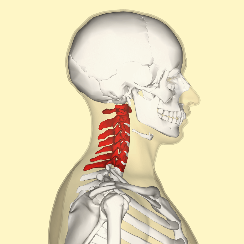
As indicated by its name, the vertebral compartment of the neck contains the cervical portion of the vertebral column. The vertebral compartment is in the posterior part of the neck. The cervical portion of the vertebral column contains the cervical vertebrae and the cartilaginous discs interposed between the cervical vertebrae. The cervical region of the spinal cord is present within the cervical vertebral column, so it is also included in the vertebral compartment of the neck.
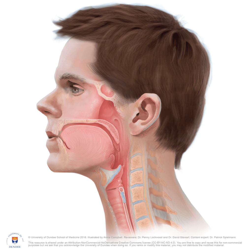
The visceral compartment of the neck is the anterior portion of the neck that contains the visceral structure of the respiratory and gastrointestinal systems. The visceral compartment also contains some endocrine glands. These structures present in the visceral compartment include the larynx, pharynx, trachea, esophagus, thyroid, and parathyroid gland.
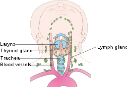
There is a pair of vascular compartments in the neck. Each vascular compartment is located on the sides of the trachea. The main content of the vascular compartments of the neck is two carotid sheaths. Each carotid sheath contains the common carotid artery and internal jugular vein. The vascular compartments of the neck also contain the vagus nerve and lymph nodes surrounding the blood vessels.
Various neck muscles attach to the inferior part of the skull, sternum, clavicles, and hyoid bone. These muscles are arranged to form two triangles in the neck; the anterior and posterior triangles. These triangles are created bilaterally, so there is a total of four triangles in the neck.
The anterior triangle of the neck is bounded medially by the midline of the neck, laterally by the anterior border of the sternocleidomastoid muscle, and superiorly by the inferior edge of the mandible. The anterior triangle contains several suprahyoid and infrahyoid muscles.
The anterior triangle’s suprahyoid muscles, which include stylohyoid, digastric, mylohyoid, and geniohyoid, are located above the hyoid bone. When these suprahyoid muscles contract, they elevate the hyoid bone. The infrahyoid muscles of the anterior triangle include omohyoid, sternohyoid, thyrohyoid, and sternothyroid. These muscles are located below the hyoid bone, and they dress the hyoid bone by their contraction.
The posterior triangle of the neck is bounded inferiority by the middle third of the clavicle, anteriorly by the posterior border of the sternocleidomastoid muscle, and posteriorly by the anterior border of the trapezius muscle. The muscles present in the posterior triangle of the neck include splenius capitis, levator scapulae, omohyoid, and scalene muscles. Some part of the sternocleidomastoid and trapezius is also present in the posterior triangle.

The neck is a transition between the head and torso of your body. The functions of the neck are mentioned in the following section.
The muscles of the neck support and hold the head at its position. But the neck muscles do not fix the head; instead, they allow the head to move up to a certain extent in different directions. This mobility and support of the head by the neck are necessary to perform routine activities.
Different structures pass through the neck. These structures include the organs of the gastrointestinal and respiratory systems. The esophagus and the trachea pass through the neck. The muscles of the neck hold these structures in their position and prevent diversions. Several nerves from the CNS also reach the organs through the neck.
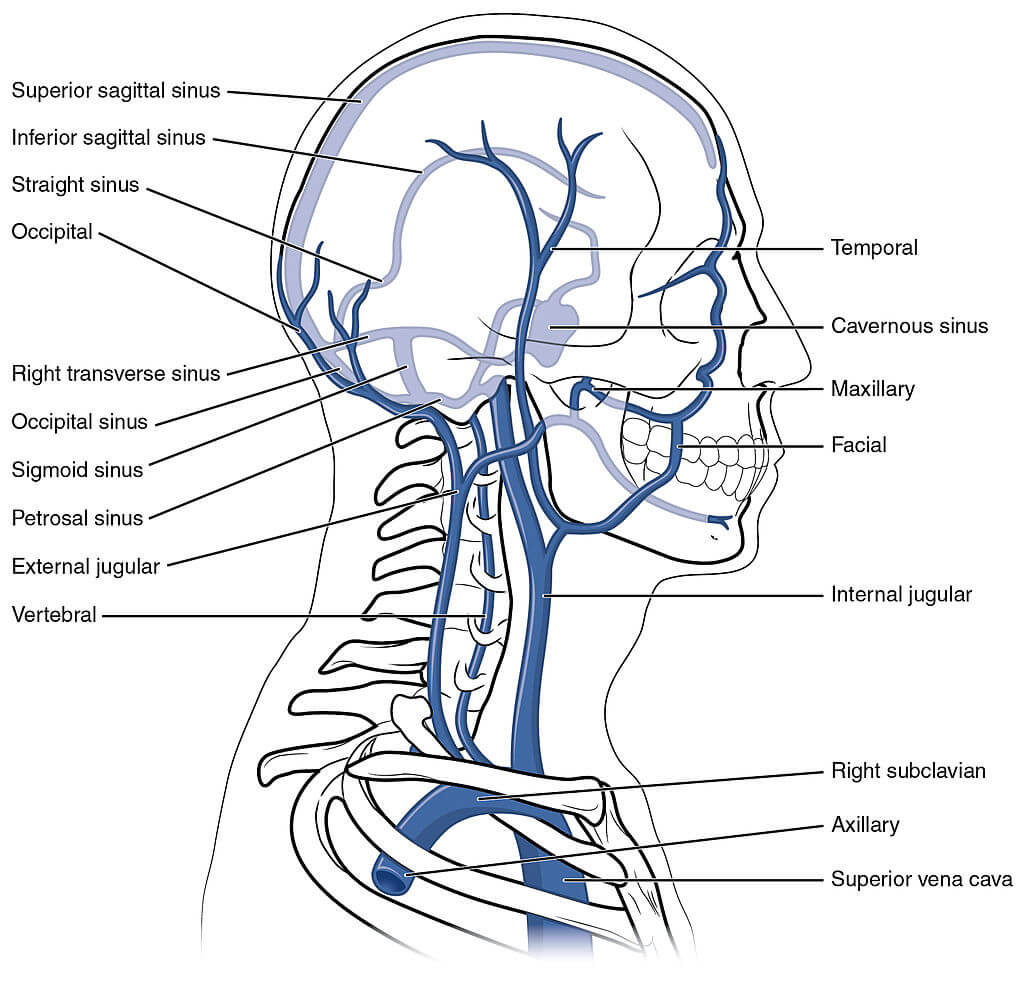
The blood supply of the brain and face is through the arteries that pass through the neck. Similarly, the brain and face venous drainage returns to the heart through veins that traverse the neck.
The blood supply of the neck is from the branches of the common carotid arteries. The right and left common carotid arteries to divide into internal and external carotid arteries, which supply blood to the neck.
The venous drainage of the neck can be divided into superficial and deep drainage. The superficial venous drainage of the neck from the subcutaneous tissue and skin occurs through the anterior and external jugular vein. The venous drainage of the deep structures of the neck occurs through the internal jugular vein.
The nerve supply of the anterior part is from the roots of cervical spinal nerves C2-C4. The nerve supply of the posterior of the neck is also from the roots of cervical spinal nerves, but in posterior parts, it is from C4-C5 instead of C2-C4. The vagus and accessory cranial nerve also traverse through the neck.
The neck has a lot of functioning in routine activities, and it bears a lot of stress. This stress on the neck can disrupt the neck, which leads most common clinical condition related to the neck area, i.e., neck pain. The most common cause of neck pain include:
Besides these conditions, some diseases or obstructions of the trachea and esophagus can affect the neck.
Note: One most important clinical significance of the neck is its rigidity in patients with meningitis. Neck rigidity is a strong lead in the diagnosis of meningitis.
The neck is the structure that connects your head with your torso. The neck is structurally divided into four compartments. These compartments vertebral compartment, visceral compartment, and two vascular compartments. The compartments have been named according to the structures they contain.
Muscles of the neck, clavicle and inferior border of the jaw form the boundaries of the triangles of the neck. These boundaries divide the neck into anterior and posterior triangles. Triangles of the neck contain muscles and vascular structures. The blood supply of the neck is from the carotid arteries, and venous drainage occurs through the jugular veins. The nerve supply of the neck is from the cervical spinal nerves.
The primary function of the neck is to provide support and motility to the head. It also acts as a safe passage for different structures. The most common clinical condition related to the neck is neck pain. Various causes of neck pain are associated with the cervical vertebral column and blood vessels passing through the neck.
1. Kohan, E. J., & Wirth, G. A. (2014). Anatomy of the neck. Clinics in plastic surgery, 41(1), 1–6. https://doi.org/10.1016/j.cps.2013.09.016
2. O’Daniel T. G. (2018). Understanding Deep Neck Anatomy and Its Clinical Relevance. Clinics in plastic surgery, 45(4), 447–454. https://doi.org/10.1016/j.cps.2018.06.011
3. Williams D. W., 3rd (1997). An imager’s guide to normal neck anatomy. Seminars in ultrasound, CT, and MR, 18(3), 157–181. https://doi.org/10.1016/s0887-2171(97)90018-4
4. Breeland, G., Aktar, A., & Patel, B. C. (2021). Anatomy, Head and Neck, Mandible. In StatPearls. StatPearls Publishing. https://pubmed.ncbi.nlm.nih.gov/30335325/
5. Kikuta S, Iwanaga J, Kusukawa J, Tubbs RS. Triangles of the neck: a review with clinical/surgical applications. Anat Cell Biol. 2019;52(2):120-127. doi:10.5115/acb.2019.52.2.120 https://www.ncbi.nlm.nih.gov/pmc/articles/PMC6624334/
The content shared in the Health Literacy Hub website is provided for informational purposes only and it is not intended to replace advice, diagnosis, or treatment offered by qualified medical professionals in your State or Country. Readers are encouraged to confirm the information provided with other sources, and to seek the advice of a qualified medical practitioner with any question they may have regarding their health. The Health Literacy Hub is not liable for any direct or indirect consequence arising from the application of the material provided.


