3D Anatomy Model
Add another dimension to your learning with fully-interactive educational male and female anatomical models.
Learning about the human anatomy has never been more fun!
Purchase
Blood is the main transporter of all the essential nutrients required for the normal functioning of our body. From oxygen to glucose, fat, and amino acids every vital requirement of cells in our body is delivered by the circulating blood. The heart, as we all know, is the main pumping organ of our body that pushes blood through our circulation without any rest.
Aorta is the first artery that arises from the heart, therefore making it one of the most important channels for blood flow. It arises directly from the heart and gives off arteries supplying all the organs of the body. Proper functioning or aorta is crucial for optimal body functioning and preventing damage.
The aorta begins at the top left ventricle, the heart’s pumping chamber. It is about a foot-long tube, with an initial diameter of one inch, and is divided into four segments. These are:
It is the first part of the aorta beginning at the aortic orifice (circular opening), roughly at the level of the inferior border of the third costal cartilage (third rib). As the name suggests, it ascends from this point, slightly sideways, and ends at the level of the sternal angle. The sternal angle, at the level of the 2nd costal cartilage, is the palpable bony prominence in the midline of your chest.
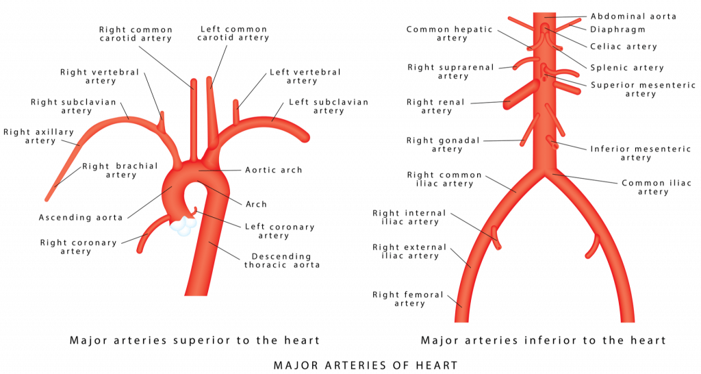
The ascending aorta gives off two branches, and despite being few in number they hold immense importance. These are the left and right coronary arteries and they supply arterial blood to the heart tissue.
The aortic arch is the second part of the aorta and begins at the branching point of the brachiocephalic trunk (branch of aorta supplying head and arms), behind the sternal angle. Curving upwards and backward to the left, the aortic arch courses in front of the tracheal bifurcation (division). It ends at the level of the fourth thoracic vertebra (T4) right after giving off the left subclavian artery, continuing as the descending aorta.
The aortic arch has a curved shape and three important branches arise from its convexity; the brachiocephalic trunk, the left common carotid artery, and the left subclavian artery.
The brachiocephalic artery supplies the right side of the head, neck, brain, and the right upper limb (shoulder and arm) through its branches.
The left common carotid artery supplies the left side of the head, neck, and brain.
The left subclavian artery supplies the left upper limb.
The Descending aorta is the largest part of the aorta. It begins as the continuation of the aortic arch. The descending aorta is further divided into the thoracic and abdominal aorta.
The thoracic aorta supplies blood to the structures in the thorax i.e., the chest cavity. It begins at the level of the T4 vertebra, coursing downwards in the posterior chamber of the thorax (posterior mediastinum). The thoracic aorta gives off several paired and unpaired arteries namely:
– Pericardial branches; supplying the pericardium (outer layer of the heart)
– Bronchial arteries; supplying bronchi and part of lungs
– Oesophageal arteries; supplying the oesophagus
– Mediastinal arteries; supplying the posterior mediastinum
– Posterior Intercostal branches; supplying the intercostal (between ribs) spaces
– Subcostal arteries; supplying the area around the lower-most ribs
– Superior Phrenic arteries; supplying the diaphragm
At the level of the T12 vertebra, the thoracic aorta ends just before passing through the aortic orifice of the diaphragm.
The abdominal aorta begins at the level of the T12 vertebra just below or at the level of the diaphragm. It terminates at the level of the L4 vertebra while bifurcating (dividing) into its terminal branches: the left and right common iliac arteries. You can visualize the bifurcation as 1.5 cm below and left to the umbilicus, on your skin.
The branches of the abdominal aorta can be divided into four main groups: the anterior (in front), lateral (to the side), dorsal (behind), and terminal branches group.
The anterior group consists of the celiac trunk, the superior mesenteric artery, and the inferior mesenteric artery.
The lateral group consists of the Suprarenal artery, the Renal artery, and the Gonadal (ovarian or testicular) arteries.
The dorsal group contains the Inferior Phrenic artery, the Lumbar arteries, and the Median Sacral arteries.
Finally, the terminal group consists of the left and right common iliac arteries.
The aorta is innervated by the aortic branch of the vagus nerve.
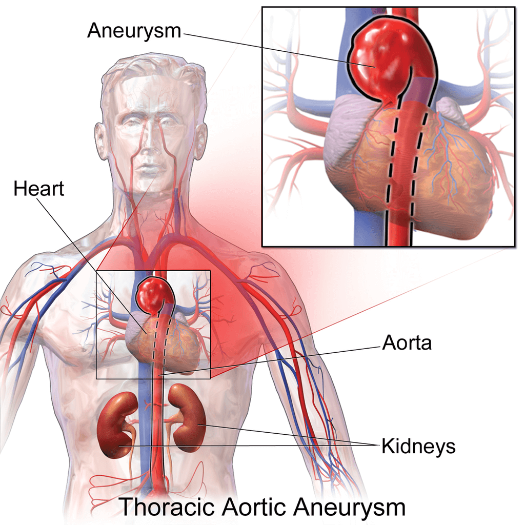
Aortic Disorders can range from aortic atherosclerosis to aortic aneurysm – a life-threatening situation. These conditions are:
Ballooning of a segment of the aorta is termed an aortic aneurysm. It is caused by a weakness in the aortic wall, that leads to expansion of that aortic segment at every stroke of the heart. Rupturing of this aneurysm can be fatal if not treated.
An aortic dissection occurs when the innermost layer of the aortic wall tears due to high blood pressure. The surging of the blood through the inner and middle layer of the aorta is called aortic dissection. There are several types of aortic dissection and sometimes it can require immediate surgery to prevent death.
This term refers to the narrowing or constriction of the aorta leading to decreased blood flow and cyanosis (bluing of the body due to decreased blood flow). It usually occurs just beyond the point where the aorta gives off branches to the head and arms.
It is a condition, caused by high serum cholesterol levels and high blood pressure, in which cholesterol plaques build up in the wall of the aorta. This can lead to a stroke i.e., interruption of blood supply to the brain.
A valve is present between the aorta and left ventricle to prevent the backflow of blood. The inability of this valve to close properly causes backflow of blood causing aortic insufficiency. This can cause the heart to pump harder eventually leading to cardiac failure.
Narrowing of the aortic valve can cause a heavy strain on the heart’s left ventricle, due to insufficient blood flowing into the aorta. This is most commonly caused by rheumatic fever, an autoimmune inflammatory condition.
Inflammation of the aorta is called aortitis. Infections and autoimmune diseases are mostly responsible for this condition.
Aortitis can be treated via different methods using medications and surgery.
The content shared on the Health Literacy Hub website is provided for informational purposes only and it is not intended to replace advice, diagnosis, or treatment offered by qualified medical professionals in your State or Country. Readers are encouraged to confirm the information provided with other sources and to seek the advice of a qualified medical practitioner with any question they may have regarding their health. The Health Literacy Hub is not liable for any direct or indirect consequence arising from the application of the material provided.


