3D Anatomy Model
Add another dimension to your learning with fully-interactive educational male and female anatomical models.
Learning about the human anatomy has never been more fun!
Purchase
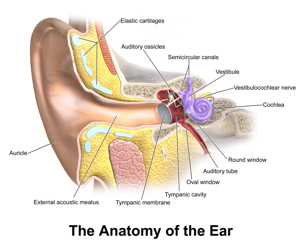
The ear is divided into three parts; external, middle, and inner ear. Different structures present in various parts of the ear have a role in hearing and maintaining the equilibrium and balance of the body. The vestibule of the ear is the central part of the bony labyrinth of the ear.
Here are some key facts about the vestibule.
1. In anatomy, the term vestibule describes a small cavity that opens into another chamber or a canal, like a porch at the entrance of a building. Various examples of vestibules exist in the human body. These include the vulvar vestibule, which is the tissue between the labia minora that surrounds the opening of the vagina (urethral and vaginal orifices). The nasal cavity also has a vestibule (nasal vestibule), which corresponds to the nostril opening, characterized by the presence of hair cells that filter foreign particles and microorganisms from the air entering your body. In the vestibule of the vagina, a type of gland known as the vestibular gland is responsible for secreting mucus that moistens the labia and vestibule. In the inner ear, the vestibule is the space between the tympanic cavity and the posterior region of the cochlea.
2. The ear vestibule is medial to the tympanic membrane, posterior to the cochlear duct, and anterior to the semicircular canal (also known as semicircular ducts).
3. The vestibule contains an otolith organ named utricle, and another known as saccule.
4. The vestibule of the ear is mainly related to the control of equilibrium and balance of your body.
5. A pathology affecting the vestibular system usually results in a loss of control over the balance and equilibrium of the body.
Let’s explore in detail the structure and functions of the ear vestibule, and the clinical relevance of this body part.
The vestibule of the ear is somewhat oval, that has a flat surface in the transverse section. The length of the vestibule is around 5mm; the width is almost. It is like a cavity within the temporal bone containing organs and nerves related to the ear’s vestibular system. In the following sections, we will review in detail the different parts of the vestibule.
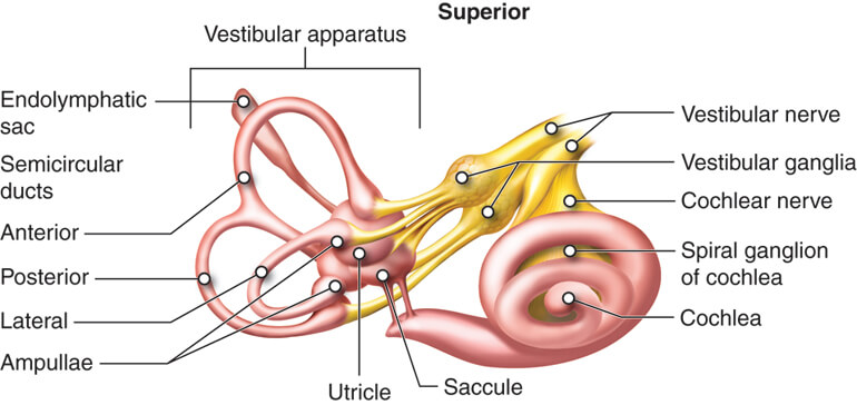
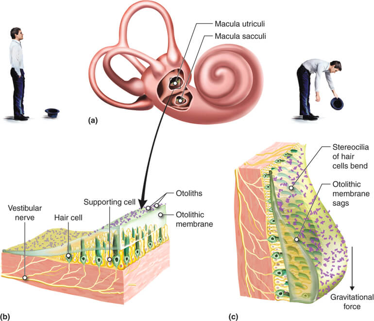
The two otolith organs found in the vestibule include the utricle and saccule. Both of these otolith organs contain sensory epithelium and macula. The macula acts as a receptor in the otolith organs of the vestibule.
The utricle lies in the posterior part of the vestibule of the inner ear. It is close to the semi-circular canals. The macula or receptor of the utricle is directed horizontally.
The saccule is the smaller and anteriorly located part of the vestibule. It has proximity to the cochlea. The macula or receptor of the saccule is positioned vertically.
The macula is found in both the utricle and saccule. It contains hair-like sensory structures, which are of two types.
Kinocilium is a single hair-like structure that is the true cilium. In a bundle of hair-like receptors, kinocilium is the longest structure and always stays erect.
The stereocilia are hair-like structures that are found as bundles. The stereocilia are not true cilia, but they also contain actin filaments. The stereocilia are flexible and shorter. According to the position of the body, they move towards or away from the adjacent kinocilium.
The hair-like sensory bundle of the macula is split into two halves by a structure called striola. The striola is an otolithic membrane separated from the sensory bundles by a layer of gelatinous material. Small calcium carbonate crystals are embedded in the striola, which are called otoconia.
The primary function of the vestibule is to send signals to your brain about equilibrium, balance, and position of your body. Varying degrees and types of signals are transferred to the brain from the vestibule according to changes in position, velocity, and equilibrium. The discussion of the functions of the individual parts of the vestibule is in the following section.
As said above, the utricle is positioned in a horizontal plane. The horizontal position of the utricle makes it sensitive to movements of the horizontal plane, such as tilting of the head. Hence, the sense of position and equilibrium of a standing person is conveyed to the brain by the utricle. The utricle also creates communication with other ear parts, allowing your brain to get more accurate sensory information about your position and equilibrium.
The position of the saccule is in the vertical plane. Hence, it has the function of detecting movements of the longitudinal plane. The movements in the longitudinal plane are made when you are lying down.
The hair cells are arranged on two sides of the midline striola as mirror images. When the hair cells from one side move towards the midline, the hair cells of the other side move away from the midline. The movement of the hair cells towards or away from striola gives the brain a gross signal of equilibrium or instability.
The main blood supply of the vestibule is from the internal auditory artery (Labyrinthine artery). The internal auditory artery may arise from the anterior and superior cerebellar artery. It may also arise from the basilar artery.
The venous drainage of the vestibule occurs through the labyrinthine vein. The labyrinthine vein subsequently opens into the sigmoid sinus or inferior petrosal sinus.
The nerve supply of the vestibule is from the 8th cranial nerve or vestibulocochlear nerve. The vestibulocochlear nerve divides into the Vestibular nerve and cochlear nerve after it enters the ear. It is the vestibular parts of the vestibulocochlear nerve that innervates the vestibule.
The clinical conditions related to the vestibule of the ear affect your sense of stability and equilibrium. These clinical conditions can affect any part of the vestibule. The common clinical conditions related to the vestibule are in the following section.
Benign Paroxysmal Positional Vertigo is a condition in which there is a sudden loss of stability. This problem occurs due to a defect in the otoconia in the macula of either of two parts of the vestibule, i.e., utricle or saccule.
Migraine Associated with Vertigo is another condition that is considered related to the vestibule. The exact cause of this condition is unknown, but it is hypothesized that there is an inappropriate signal interpretation by the brainstem conveyed by the utricle and saccule.
Some other disorders are not directly related to the vestibule but are closely related to the functional impairment of the otolith organs. These disorders include Meniere’s disease, labyrinthitis, and vestibular neuritis.
The vestibule is the part of the inner ear related to your body’s control of balance and equilibrium. It has otolith organs named utricle and saccule that senses changes in position and equilibrium. The otolith organs have macula, which contain hair-like cells. The movement of these hair-like cells gives a signal about the position and equilibrium of the body.
The utricle is positioned horizontally and senses the horizontal plane’s movements, such as tilting the head. Contrarily, the saccule is positioned vertically, and it detects body movements in the longitudinal plane. The blood supply and venous drainage of the vestibule are through the labyrinthine vessels. The nerve supply of the vestibule is from the vestibular part of the 8th cranial nerve. Any damage problem to the vestibule results in vertigo.
1: Bruss DM, Shohet JA. Neuroanatomy, Ear. [Updated 2020 Jul 31]. In: StatPearls [Internet]. Treasure Island (FL): StatPearls Publishing; 2021 Jan-. Available from: https://www.ncbi.nlm.nih.gov/books/NBK551658/
2: DELATTRE, A., & FENART, R. (1961). Journal des sciences medicales de Lille, 79, 100–104. https://pubmed.ncbi.nlm.nih.gov/13721491/
3: Ciuman R. R. (2009). Stria vascularis and vestibular dark cells: characterization of main structures responsible for inner-ear homeostasis, and their pathophysiological relations. The Journal of laryngology and otology, 123(2), 151–162. https://doi.org/10.1017/S0022215108002624
4: Ekdale E. G. (2016). Form and function of the mammalian inner ear. Journal of anatomy, 228(2), 324–337. https://doi.org/10.1111/joa.12308
5: Fík, Z., & Bouček, J. (2019). Inner ear disorders. Onemocnění vnitřního ucha. Casopis lekaru ceskych, 158(6), 216–220. https://pubmed.ncbi.nlm.nih.gov/31931577/
The content shared in the Health Literacy Hub website is provided for informational purposes only and it is not intended to replace advice, diagnosis, or treatment offered by qualified medical professionals in your State or Country. Readers are encouraged to confirm the information provided with other sources, and to seek the advice of a qualified medical practitioner with any question they may have regarding their health. The Health Literacy Hub is not liable for any direct or indirect consequence arising from the application of the material provided.


