Overview
A PET scan or a positron emission tomography scan is an advanced imaging test that can detect several abnormalities related to the biochemistry of the body. PET scan is a mixture of nuclear medicine and biochemical testing. It basically uses a nuclear agent (similar to the contrast agent of CT scan) to measure the metabolic activity of a particular organ or tissue in our body.
A PET scan is very helpful in identifying cancers or tumours, heart diseases, and brain abnormalities. It can often detect diseases in earlier stages before it shows up on other imaging tests like CT (computed tomography) scan or MRI (magnetic resonance imaging). In this article, we will go through the mechanism, procedure, and uses of positron emission tomography (PET) scan.
Indications
As mentioned above, PET scan is a useful technique for detecting and diagnosing several abnormal body conditions. Your doctor will use the scans to diagnose your condition and prescribe prompt treatment. Certain indications for a PET scan are:
- Cancer or malignancies – cancer is a serious disease that is often lethal if not detected in its early stages. PET scan is used for the diagnosis of malignancy, staging of cancer, tumour characterisation, treatment response assessment, surveillance of the disease.
- Infection or inflammation – PET scans are useful in diagnosing chronic conditions like rheumatic diseases, vasculitis, etc.
- Heart disease – PET scans can help visualise areas of decreased blood flow in the heart or clogged arteries.
- Brain disorders – PET scans are ordered often in the diagnosis and management of certain brain conditions like Alzheimer’s disease, brain tumours, seizures.
Risks of the Procedure
PET scan requires a nuclear medicine or tracer (like a contrast agent) to be injected into your body intravenously (into your vein). This tracer is a radioactive substance that can have harmful effects on your body. However, the intensity of radiation that you get exposed to is very low and is not that harmful. You might experience a few side effects like:
- May harm your unborn baby if you’re pregnant
- May expose your breastfeeding child to radiation through your breast milk
- Rarely, cause an allergic reaction
- May traumatise claustrophobic patients
You should discuss the risks and benefits of getting a PET scan with your doctor or a nuclear medicine specialist.
Preparation
Your physician will provide you with the general instructions and preparation measures for the PET scan. You would need to follow some simple precautions before your test:
- Let your doctor know if you had any allergies in the past, especially during a test
- If you have any medical conditions such as hypertension, phobia (e.g., claustrophobia), or any recent sickness
- If you’re pregnant or breastfeeding your child
- You would have to empty your bladder before the PET scan
- You will have to change into a hospital gown
- Generally, you should avoid strenuous exercise for a couple of days before your PET scan
- Injecting the radioactive drug or tracer – the doctor or technician will administer the tracer intravenously through a vein in your arm or hand. You would need to relax for 30-60 minutes until the drugs get absorbed into your body properly. You might feel something cold going up your arm during tracer administration.
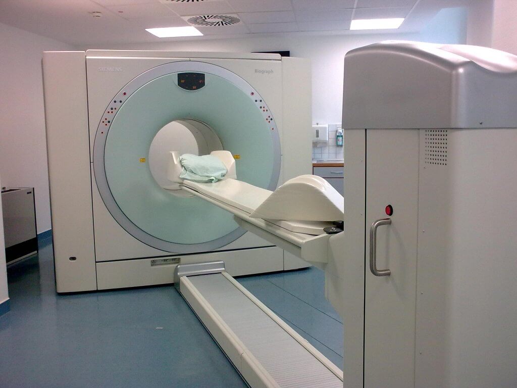
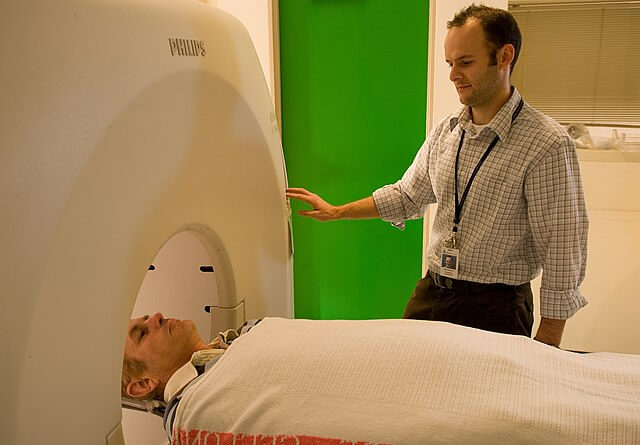
A PET scanner is a large doughnut-like machine surrounding the lying table. It is similar to CT or MRI scanners.
Procedure
During the procedure, you lie on a flat, narrow, and padded table. You might be strapped throughout the procedure in order to limit physical movement. This table slides in and out of the large doughnut-like hole of the scanner.
PET scans are often combined with a CT or MRI, i.e., a PET-CT scan or PET-MRI scan. A PET-CT scan usually takes about 30 minutes while a PET-MRI scan takes about 45 or so minutes.
The machine or scanner is moving around at a fast pace, so it makes a lot of buzzing and clicking sounds which are completely normal.
The scan is completely painless. However, you may feel some anxiety laying down in a closed space for 30-60 minutes. The technician will be in the room next to you observing you and hearing you. You can tell them if you feel uncomfortable or anxious.
Patient Recovery
The PET scan will not cause you any disturbance in your daily life. After the test, you can go back home and carry on your routine work. You should drink plenty of water in order to excrete the tracer (nuclear medicine). You should follow your doctor’s instructions if they advise you otherwise.
Results
A radiologist (specialised doctors to do imaging tests and interpret them) will look at your scan results and interpret them. They will then tell your doctor about the findings. The doctors might compare your results from this scan to any previous tests like CT or MRI, to evaluate the results. This process usually takes 24 – 48 hours.
The results of a PET scan can be used to:
- Diagnose cancer or malignant disease
- Check the progression of your disease
- Evaluate the effectiveness of the treatment you’re going through
In case of certain findings, your doctor may prescribe another PET scan or a different type of PET scan according to your results.
- Positron emission tomography — Computed tomography (PET/CT). Radiological Society of North America. https://www.radiologyinfo.org/en/info.cfm?pg=PET. Accessed April 6, 2021.
- What is PET? Society of Nuclear Medicine and Molecular Imaging. https://www.snmmi.org/AboutSNMMI/Content.aspx?ItemNumber=5649. Accessed April 6, 2021.
- Umterrainer M, et al. Recent advances of PET imaging in clinical radiation oncology. Radiation Oncology. 2020; doi:10.1186/s13014-020-01519-1.
- Adam A, et al., eds. Adrenal imaging. In: Grainger and Allison’s Diagnostic Radiology. 7th ed. Elsevier; 2021. https://www.clinicalkey.com. Accessed April 6, 2021.
- ACR-SPR practice parameter for performing FDG-PET/CT in oncology. American College of Radiology. https://www.acr.org/Clinical-Resources/Practice-Parameters-and-Technical-Standards/Practice-Parameters-by-Modality. Accessed April 6, 2021.
- Cervical cancer. Radiological Society of North America. https://www.radiologyinfo.org/en/info.cfm?pg=cervicalcancer. Accessed April 8, 2021.
- Morrow ES. Allscripts EPSi. Mayo Clinic. April 6, 2021.
- Collins DA (expert opinion). Mayo Clinic. April 24, 2021
The content shared in the Health Literacy Hub website is provided for informational purposes only and it is not intended to replace advice, diagnosis, or treatment offered by qualified medical professionals in your State or Country. Readers are encouraged to confirm the information provided with other sources, and to seek the advice of a qualified medical practitioner with any question they may have regarding their health. The Health Literacy Hub is not liable for any direct or indirect consequence arising from the application of the material provided.