Overview
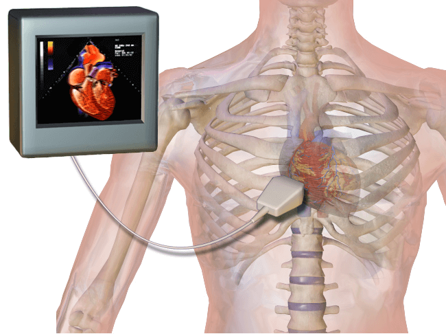
An echocardiogram uses sound waves to produce an image of an organ. It uses sound waves to perform images and essentially an ultrasound. Echocardiograms or an echo are used for imaging of the heart. By being able to see the heart work in real-time, your physician is able to decipher whether or not the blood flow and pumping of the heart indicate any cardiac (heart) disease.
Given the circumstances, you may require one of many types of echocardiograms. Each echocardiogram is slightly different from the other and involves few specific risks if not at all. In this article, we will go through some details about echocardiograms.
Indications
Your physician may suggest getting an echocardiogram if there are any problems with the heart. These problems could be detrimental and therefore getting an echo becomes imperative. Some issues that an echocardiogram can detect are:
- Problems with the valves and chambers of the heart
- Heart problems being the root of shortness of breath and angina (chest pain)
- Congenital heart birth defects – Birth defects of the heart that have a genetic cause and are present since birth.
- Weakened heart muscles
- Blood clots in the vessels and heart
- Tumors of the heart
- Enlargement of heart chambers
- Effect of medical treatment
Risks of Procedure
There are little to no risks involved with standard echocardiograms. The probe or transducer will be held firmly against your chest and may cause a sense of pressure. This is done so that the best images may be performed. If the echocardiogram is performed in an area such as the throat some soreness may last for a couple of hours. In rare events, the transducer may scrape the skin. Oxygen saturation is actively monitored to find indications of distress from sedation.
Sometimes an echocardiogram is performed during a period of stress. A situation that can trigger stress is strenuous exercise. By being in a state of stress, an issue within the heart becomes visible which would otherwise not be seen if the echo was performed at rest. Extremely serious consequences like heart attacks are unheard of.
Patient Preparation
Special preparations like avoiding eating/drinking or medications are not necessary for some echocardiograms. In other echocardiograms, you will not be able to drive back home due to medications.
An echocardiogram can be performed both in the hospital and the doctor’s office. Most echoes only take a maximum of an hour. The procedure follows the following steps for a transthoracic echocardiogram:
- You will undress from the waist up.
- The technician will attach electrodes to your body to detect the heart’s electrical impulses.
- A gel will be applied onto the transducer and move over your chest to detect the echoes from your heart.
- The technician may advise you to change the way you breathe and/or rollover to your left side.
However, in a transesophageal echocardiogram you will:
- Have your throat numbed.
- Given a sedative to help relax.
- Have a transducer inserted into your throat to get clear images of your heart while in the esophagus.
- Be asked to stay for few hours to monitor you.
Procedure
There are several types of echocardiograms. The one you will be required to take depends on what information your physician requires.
Transthoracic Echocardiogram
- A sonographer (ultrasound technician) spreads spread a gel on a prob-like device called a transducer.
- The sonographer then firmly presses the transducer against your skin to provide the clearest image of your heart with the ultrasound beam.
- As the sound wave is passed through, the wave bounces back and echoes.
- The transducer records the echoes.
- A computer to which the transducer is attached converts the echo into moving images.
In the case that either your lungs or ribs may obscure the heart, an enhancing agent may be given to you intravenously. The enhancing agents are tolerable and help enhance the images of the heart on the computer.
If the image is still not detailed enough, your physician may order a transesophageal echocardiogram
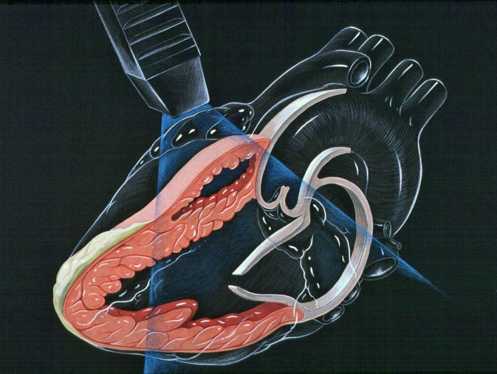
Transesophageal Echocardiogram
- Your throat is numbed by medication to avoid uncomfortableness.
- A tube attached to the transducer is inserted down your esophagus near your stomach.
- The echo then records the sound waves bounced from the heart.
- These echoes are then converted into images on the computer.
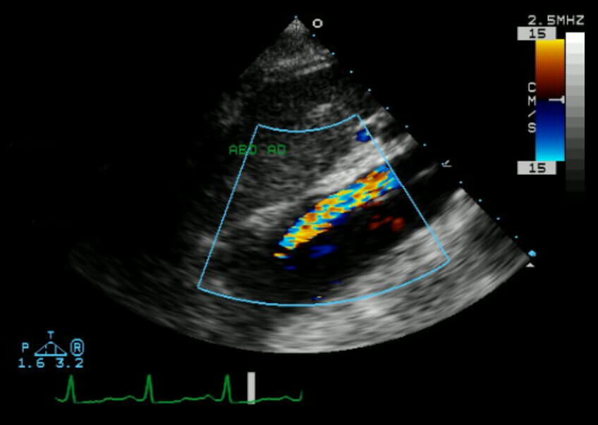
Doppler Echocardiogram
In a Doppler echocardiogram, your physician can determine the speed and direction by which the blood is moving in your circulatory system. The blood cells actually change the pitch of the sound wave once it bounces off. This change is picked up by the transducer. This echo can be used in arteries as well to make sure there are no blood movement issues that most traditional ultrasounds cannot pick up. And finally, the stress and fetal echocardiogram are taken when in stress and while in the mother’s womb respectively.
Patient Recovery
Most people can go back to their normal daily activities without any issues. If your results seem abnormal you will most probably be referred to a cardiologist to discuss the abnormal test in more detail.
Outcomes
The echocardiogram may show:
- Change in heart size – damaged and weakened heart valves, hypertension, and other comorbidities may cause the chamber to become enlarged or thickened.
- Damage to heart muscles – Areas of the heart that are damaged move less and can indicate occurrences such as heart attacks.
- Valve problems – issues in the valves may cause the valve to become too enclosed or not closed enough which leads to leakage.
- Heart defects – Most defects in the heart are congenital and are usually indicated by abnormal connections of the heart and blood vessels.
- Bonow RO, et al., eds. Echocardiography. In: Braunwald’s Heart Disease: A Textbook of Cardiovascular Medicine. 11th ed. Philadelphia, Pa.: Saunders Elsevier; 2019. https://www.clinicalkey.com. Accessed Sept. 2, 2018.
- Goldman L, et al., eds. Echocardiography. In: Goldman-Cecil Medicine. 25th ed. Philadelphia, Pa.: Saunders Elsevier; 2016. https://www.clinicalkey.com. Accessed Sept. 2, 2018.
- Zitelli BJ, et al. Cardiography. In: Zitelli and Davis’ Atlas of Pediatric Physical Diagnosis. 7th ed. Philadelphia, Pa.: Elsevier; 2018. https://www.clinicalkey.com. Accessed Sept. 2, 2018.
- Echocardiography. National Heart, Lung, and Blood Institute. https://www.nhlbi.nih.gov/health-topics/echocardiography. Accessed Sept. 2, 2018.
- Mankad R (expert opinion). Mayo Clinic, Rochester, Minn. Sept. 26, 2018.
- https://www.webmd.com/heart-disease/guide/diagnosing-echocardiogram
The content shared in the Health Literacy Hub website is provided for informational purposes only and it is not intended to replace advice, diagnosis, or treatment offered by qualified medical professionals in your State or Country. Readers are encouraged to confirm the information provided with other sources, and to seek the advice of a qualified medical practitioner with any question they may have regarding their health. The Health Literacy Hub is not liable for any direct or indirect consequence arising from the application of the material provided.