Eye examination involves a series of tests that are performed to evaluate your vision and to check your eyes for any diseases or complications. The eye exam requires the use of different instruments and procedures for the evaluation of eyes.
In this article, we will focus on some of the more common and clinically important eye tests. Each of these tests evaluates several aspects of your eyes and can be useful in determining different types of diseases i.e., an ophthalmology exam.
Indications
Most eye tests are indicated if you’re having some problem in your eyes or with your vision (seeing things). Some of the common indications for an eye exam are:
- Acute eye injury
- Eye pain
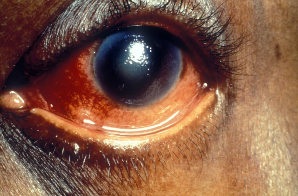
- Acute red eye
- Sudden vision loss
- Eye discharge
- Double vision (diplopia)
- Blurry vision
- Light flashes
- Chronic diseases like diabetes, multiple sclerosis
- As a general check-up – eye tests are performed at different ages throughout your life by your doctor in order to make sure that your eyes are functioning properly. These routine check-ups are done for:
- Children – your paediatrician will examine your child’s eye for healthy eye development and look for any problems like lazy eyes or cross-eye, etc. For children of age 3 to 5 your doctor may check your child’s eyes for vision and eye development.
- Adults – it is recommended that you get an eye exam around the age of 40 regardless of having any problems or not. This is the age when some of the common eye diseases start to appear and cause problems, so they can be treated as early as possible.
- If you’re 60 years or older, you are recommended to get an eye examination every year or two. If you wear glasses or contact lenses, have a family history of eye problems, or take certain medicines that are toxic to the eyes.
Risks
The usual eye examination is superficial (non-penetrating) and does not cause any harm to the eyes or any structure of the body.
Most detailed eye examinations require the use of some pupil dilating eye drops. This eye drops usually don’t cause any harm and you would just need to wear your sunglasses after your exam. However, some adverse effects that can be caused by the use of dilating eye drops include:
- An attack of narrow-angle glaucoma (especially if you’re old)
- Dizziness
- Flushing and dry mouth
- Nausea or vomiting
During some procedures, your doctor may use some anaesthetic drops to numb your eyes. This local anaesthetic may make you feel numb around your eye after your exam.
Patient Preparation
An ophthalmologist will completely examine your eyes, i.e., your visual acuity or diagnosing complex diseases, and even perform surgery if needed.
You should consider taking the following precautions:
- If you wear glasses or contact lenses, you should bring them along with you so your doctor can make sure they are still the best fit for you.
- You should bring sunglasses with you to wear them after your exam, as some procedure might make your eyes sensitive to the sunlight.
- You should also consider someone else driving you back home.
Procedure
An eye exam will consist of various procedures and it might take up to an hour depending on the health of your eyes. We will discuss some important tests in detail, momentarily.
A usual examination almost always starts with some questionnaire. If it is your first eye exam your doctor (ophthalmologist) might ask you a couple of questions including:
- Are you having any eye problems and since when?
- Did you have any eye problems in the past?
- Do you wear glasses or contact lenses? Are you satisfied with them?
- Were you born prematurely?
- Did you have any previous eye treatment?
- Did you ever have any significant problems in your eyes, i.e., itching, reddening, etc?
- Is there anyone in your family with eye problems or had a specific eye disease?
- Do you have any other chronic diseases like diabetes or heart disease?
- If you use any drugs or smoke tobacco?
We will now go through some routinely done exams of the eyes.
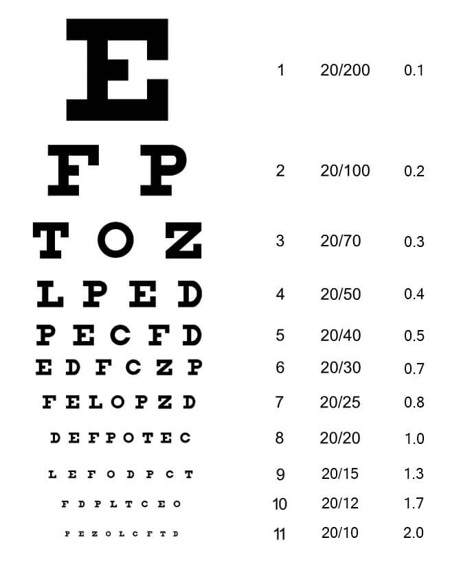
Visual acuity test
This is a very common test that your doctor will use to check how you can see. Your doctor will make you sit at a certain distance from a chart and then ask you to read that chart. It is a fairly simple test, with no risks or complications. Each of your eyes will be tested separately.
Your near vision would also be tested using charts held at reading distances.
Refraction assessment
This assessment is used to determine the best refractory power for your contact lenses or your glasses. Your doctor will determine the power that will help you have the sharpest vision.
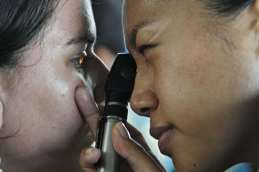
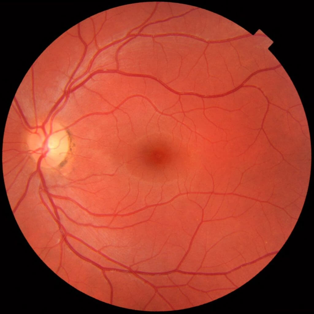
Retinal examination
This is the main examination done by an ophthalmologist and it is also called ophthalmoscopy or fundoscopy. Your doctor evaluates your eyes thoroughly including your retina (back of your eye), the optic disk, and the retinal blood vessels.
The doctor will use eye drops to dilate your pupils. Pupils allow the amount of light that enters your eyes. Dilated pupils allow your doctor to see the inside of your eyes in detail.
You will be asked to lay down on a flat or recline against a chair.
The dilating eyedrops used before the procedure can sometimes have some adverse reactions as described previously.
Fundoscopy can be done in two ways. After administering the drops your doctor may use the:
- Direct method – your doctor will shine a beam of light directly through your pupil to see the back of your eye. Both of your eyes would be tested.
- Indirect method – this is a very useful method to assess your retina as it gives a three-dimensional view of the structures present in your eyes. Your doctor will use a condensing lens and a bright light mounted on their forehead.
- Slit–lamp ophthalmoscopy – you will be sitting in a chair with your chin placed on the instrument placed in front of you and your forehead will be also kept in place so you don’t move. Your doctor will then use a microscope part of the slit-lamp and some other tiny lens to visualise your retina. This procedure allows for even higher magnification of the inner structures of the eye.
Ophthalmoscopy may be used to detect certain abnormalities within your eye, especially the ones related to the retina. Some problems that may be detected by this procedure are:
- Retinal inflammation e.g., CMV retinitis Diabetic retinopathy
- Glaucoma
- Age-related macular degeneration causing loss of sharp, central vision
- Melanoma of the eyes
- Problems associated with the optic nerve
- Any problem related to the blood vessels
- Retinal detachment
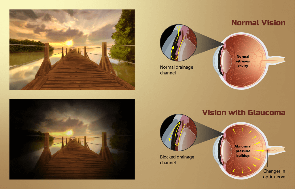
Tests for screening Intraocular Pressure (Glaucoma)
Intraocular pressure refers to the pressure in the eye due to the presence of the aqueous humour. Increased intraocular pressure can result in a condition called glaucoma which if not treated can cause blindness. It is a relatively common condition and has various causes.
We will briefly go through some of the tests used to check the intraocular pressure.
Applanation tonometry – this test determines the force needed to flatten a part of your cornea. Your doctor will give you fluorescein-containing eyedrops (used in slit-lamp as well) and some eye drops for anaesthesia.
- Using the tonometer, your doctor will touch the cornea and measure the pressure within your eye. You won’t feel any pain due to the effect of the anaesthetic.
- Non-contact tonometry – in this procedure a puff of air is used to estimate the intraocular pressure of your eyes. No anaesthetic is used in this test as your eyes do not come in contact with any instruments. You’ll feel a pulse-like feeling due to the puff of air.
- Ophthalmoscopy or fundoscopy (discussed above)
Detecting glaucoma and prompt treatment can avoid complications like vision loss and impaired vision.
Patient Recovery
After your eye examination, your doctor will discuss the results of your exam. You might be advised to follow some protective and preventive measures in order to protect your eyes and increase your visual longevity.
Outcomes
The eye exam will determine whether:
- Your eyes are normal and functioning properly and you don’t need treatment
- You need visual corrections i.e., glasses or contact lenses – in this case, your doctor will give you a prescription number.
- You need any medications for certain diseases like glaucoma
- You need surgery
- You would need more exams to rule in or rule out other diseases.
- See the full picture of your health with an annual comprehensive eye exam. American Optometric Association. https://www.aoa.org/healthy-eyes/caring-for-your-eyes/full-picture-of-eye-health?sso=y. Accessed Jan. 8, 2021.
- Eye exam and vision testing basics. American Academy of Ophthalmology. https://www.aao.org/eye-health/tips-prevention/eye-exams-101. Accessed Jan. 8, 2021.
- Difference between an ophthalmologist, optometrist and optician. American Association for Pediatric Ophthalmology and Strabismus. https://aapos.org/glossary/difference-between-an-ophthalmologist-optometrist-and-optician. Accessed Jan. 8, 2021.
- Get an eye disease screening at 40. American Academy of Ophthalmology. https://www.aao.org/eye-health/tips-prevention/screening. Accessed Jan. 8, 2021.
- Eye screening for children. American Academy of Ophthalmology. https://www.aao.org/eye-health/tips-prevention/children-eye-screening. Accessed Jan. 11, 2021.
- Yanoff M, et al., eds. Examination of ocular alignment and eye movements. In: Ophthalmology. 5th ed. Elsevier; 2019. https://www.clinicalkey.com. Accessed Jan. 8, 2021.
- AskMayoExpert. Glaucoma (adult). Mayo Clinic; 2019.
- Vision screening. American Association for Pediatric Ophthalmology and Strabismus. https://aapos.org/glossary/vision-screening-description. Accessed Jan. 8, 2021.
- Evaluation of the ophthalmologic patient. Merck Manual Professional Version. https://www.merckmanuals.com/professional/eye-disorders/approach-to-the-ophthalmologic-patient/evaluation-of-the-ophthalmologic-patient. Accessed Jan. 11, 2021.
- Get a dilated eye exam. National Eye Institute. https://www.nei.nih.gov/learn-about-eye-health/healthy-vision/get-dilated-eye-exam. Accessed Jan. 11, 2021.
- Atebara NH, Miller D, Thall EH. Ophthalmic instruments. In: Yanoff M, Duker JS, eds. Ophthalmology. 5th ed. Philadelphia, PA: Elsevier; 2019:chap 2.5.
- Ball JW, Dains JE, Flynn JA, Solomon BS, Stewart RW. Eyes. In: Ball JW, Dains JE, Flynn JA, Solomon BS, Stewart RW, eds. Seidel’s Guide to Physical Examination. 9th ed. St Louis, MO: Elsevier; 2019:chap 12.
- Chuck RS, Dunn SP, Flaxel CJ; American Academy of Ophthalmology Preferred Practice Pattern Committee, et al. Comprehensive adult medical eye evaluation preferred practice pattern. Ophth
The content shared in the Health Literacy Hub website is provided for informational purposes only and it is not intended to replace advice, diagnosis, or treatment offered by qualified medical professionals in your State or Country. Readers are encouraged to confirm the information provided with other sources, and to seek the advice of a qualified medical practitioner with any question they may have regarding their health. The Health Literacy Hub is not liable for any direct or indirect consequence arising from the application of the material provided.