Table of Contents
hide
Overview
An X-ray is one of the most commonly used imaging tests used to produce images of specific body tissues. X-rays are used for a wide variety of clinical scenarios but they are of special importance in assessing the conditions of your bones.
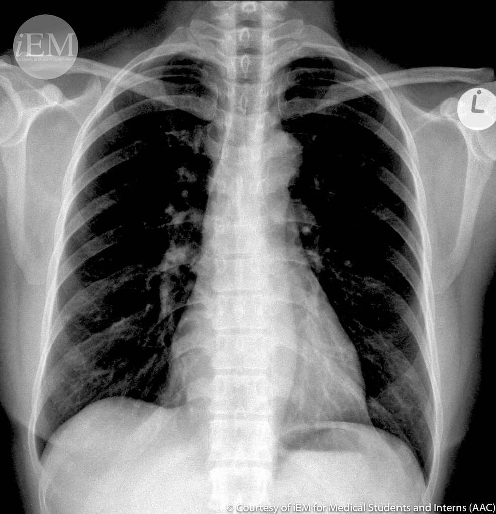
The mechanism by X-ray image is formed is fairly simple. X-ray beams are thrown at your body and these beams tend to cross your body like any other radiations. However, highly dense (compact) material likes bones and metals absorb these rays (beams). Different tissues in your body having different densities makes up the basis of an X-ray image. The bones (dense material) show up as white on an X-ray image, while other soft tissues (less dense) appear black. This black colour appears due to a lack of absorption of beams. Normal lungs appear black due to the presence of air in them. Muscle and fat tissues appear somewhat gray on an X-ray image.
In some types of X-rays, a contrast medium like iodine or barium may be used. This helps highlight a specific part of your body and gives a better image than the normal X-ray scans. In this article, we will learn about X-ray tests and some general details about X-rays.
Indications
Being a rapid and quick imaging test (done within minutes) X-rays are used to diagnose multiple different diseases. Some of the common indications for X-rays are:
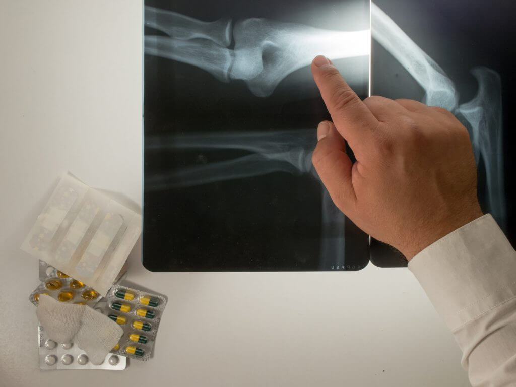
- Trauma or accident – most of the fractures of bones show up clearly on x-rays are easily diagnosed
- Chronic pain in bones which is refractory to pain killers – infections of bones and teeth can also be seen on x-rays.
- Arthritis – refers to inflammation of joints or part of your joint. Doctors widely prescribe x-rays if there is suspicion of arthritis
- Dental pain or discomfort – dentists can use to assess if there is any decay, cavities, an infection can be seen on x-rays
- Weakening of bones or recurrent fractures
- Women, especially with multiple babies and post-menopausal
- Chronic pain around a joint or pain accompanied with weight loss etc. – bone cancers are one of the indications and findings of x-rays.
- Chest or upper respiratory infection – chest X-ray has been very useful in diagnosing infections of the respiratory system e.g., pneumonia in the lungs
- Abnormalities of breast i.e., blood or pus discharge from the nipple
- If you have cardiac symptoms i.e., chest pain and angina – chest x-rays are useful in diagnosing conditions like dilated cardiomyopathies or cardiac tamponade.
- Digestive tract or swallowing problems e.g., regurgitation of food and vomit – x-rays (using contrast like barium) can reveal obstructions in the stomach or the oesophagus.
Risks
X-rays are a form of radiation. Radiations do carry a threat of inducing abnormal changes in body tissues called dysplasia or even cancer. The amount or intensity of radiation that your body gets exposed to depends on the tissue or organs being examined. Generally, the amount of radiation used in X-rays is very low and safe for most people.
The sensitivity to x-rays may depend on certain factors e.g., age, pregnancy, or a disease. In most cases, the benefits outweigh the risks of x-rays.
You should inform your doctor if you are pregnant or have a previous history of allergic reaction to some x-ray scan.
As mentioned above some X-rays use contrast medium which may cause some side effects like:
- Warmth or flushing of the skin
- Metallic taste
- Nausea or light-headedness
- Itching or allergic reaction
- Rarely, anaphylactic shock or cardiac problems
You should discuss these risks before getting your x-ray done.
Preparation
Preparation of an X-ray is fairly simple except if you’re prescribed a contrast x-ray. You would require to:
- Change into a hospital gown preferably
- Avoid wearing pins, or any metallic material
- Get the contrast medium – the contrast medium is usually barium or iodine. This can be given to you in the form of a liquid to swallow, an injection, or through an enema (through the rectum).
Procedure
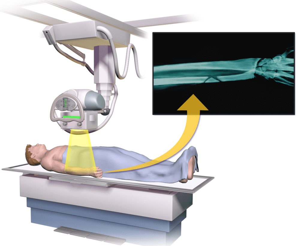
X-rays machines produce a safe level of radiation that passes through your body and the shadows get printed on a specialised plate (the black sheet). The technician will adjust the x-ray machine according to the view required. You have to remain still until the x-ray is done to avoid blurring the image.
It usually takes a couple of minutes for simple x-rays. However, it can take more time if you’re getting a contrast x-ray.
If your child is getting an X-ray, you may be advised to stay with your child in order to keep them still. If your child is moving, the x-rays might have to repeat so it is preferable to hold your child still. Sometimes, restraints of belts may be used to keep the child still.
Patient Recovery
If you had a simple x-ray you can just change back into your normal clothes and carry on with your day. Routine x-rays usually do not cause any side effects or problems.
If you were given a contrast medium, you would need to stay with the healthcare team for a little while until they make sure you’re not having any side effects. You should drink plenty of fluid for a day or two in order to clear the contrast from your body.
Results
X-rays are digitally saved on computers and the results can be visible in minutes. A radiologist or your specialised doctor will look at these image(s) and interpret them. Usually, you get the results within some hours, and interpretation of your results is done in around 24 hours.
In case of any emergencies like accidents or respiratory insufficiency, X-ray results can be made available within minutes to your doctor.
- X-rays. National Institute of Biomedical Imaging and Bioengineering (NIBIB). https://www.nibib.nih.gov/science-education/science-topics/x-rays. Accessed Dec. 7, 2019.
- Bone X-ray (radiography). Radiological Society of North America. https://www.radiologyinfo.org/en/info.cfm?PG=bonerad. Accessed Dec. 7, 2019.
- Patient safety: Contrast materials. Radiological Society of North America. https://www.radiologyinfo.org/en/info.cfm?pg=safety-contrast. Accessed Dec. 7, 2019.
- Panoramic dental X-ray. Radiological Society of North America. https://www.radiologyinfo.org/en/info.cfm?pg=panoramic-xray. Accessed Dec. 7, 2019.
- Bone mass measurement: What the numbers mean. NIH Osteoporosis and Related Bone Diseases National Resource Center. https://www.bones.nih.gov/health-info/bone/bone-health/bone-mass-measure. Accessed Dec. 7, 2019.
- Lee CI, et al. Radiation-related risks of imaging studies. https://www.uptodate.com/contents/search. Accessed Dec. 7, 2019.
- X-ray (radiography) — Chest. Radiological Society of North America. https://www.radiologyinfo.org/en/info.cfm?pg=chestrad. Accessed Dec. 7, 2019.
- Mammograms. National Cancer Institute. https://www.cancer.gov/types/breast/mammograms-fact-sheet. Accessed Dec. 7, 2019.
- Children’s (pediatric) X-ray (radiography). Radiological Society of North America. https://www.radiologyinfo.org/en/info.cfm?pg=pediatric-xray. Accessed Dec. 7, 2019.
- Zitelli BJ, et al., eds. Fundamentals of pediatric radiology. Zitelli and Davis’ Atlas of Pediatric Physical Diagnosis. 7th ed. Elsevier; 2018. https://www.clinicalkey.com. Accessed Dec. 7, 2019.
- Litin SC (expert opinion). Mayo Clinic. Dec. 8, 2019.
The content shared in the Health Literacy Hub website is provided for informational purposes only and it is not intended to replace advice, diagnosis, or treatment offered by qualified medical professionals in your State or Country. Readers are encouraged to confirm the information provided with other sources, and to seek the advice of a qualified medical practitioner with any question they may have regarding their health. The Health Literacy Hub is not liable for any direct or indirect consequence arising from the application of the material provided.