Model Anatomi 3D
Tambahkan dimensi lain ke pembelajaran Anda dengan model anatomi pria dan wanita pendidikan yang sepenuhnya interaktif.
Belajar tentang anatomi manusia tidak pernah semenyenangkan ini!
Pembelian
Sistem pencernaan memungkinkan kita memecah makanan yang kita makan untuk mendapatkan energi dan gizi. Biasanya dibagi menjadi saluran pencernaan (juga disebut saluran GI atau saluran pencernaan), hati, pankreas, dan kantong empedu. Saluran GI adalah serangkaian organ berongga yang bergabung dalam tabung yang panjang dan memutar dari mulut ke anus. Organ berongga yang menyusun saluran GI adalah mulut, kerongkongan, perut, usus kecil, usus besar, dan anus.
Organ-organ ini bergabung untuk melakukan enam tugas: menelan, mengeluarkan, mengeluarkan, pencernaan, penyerapan, dan buang air besar.
Fungsi-fungsi vital ini diperlukan untuk menjaga homeostasis yang sehat (pemeliharaan lingkungan internal yang konstan) dan untuk fungsi optimal tubuh manusia.
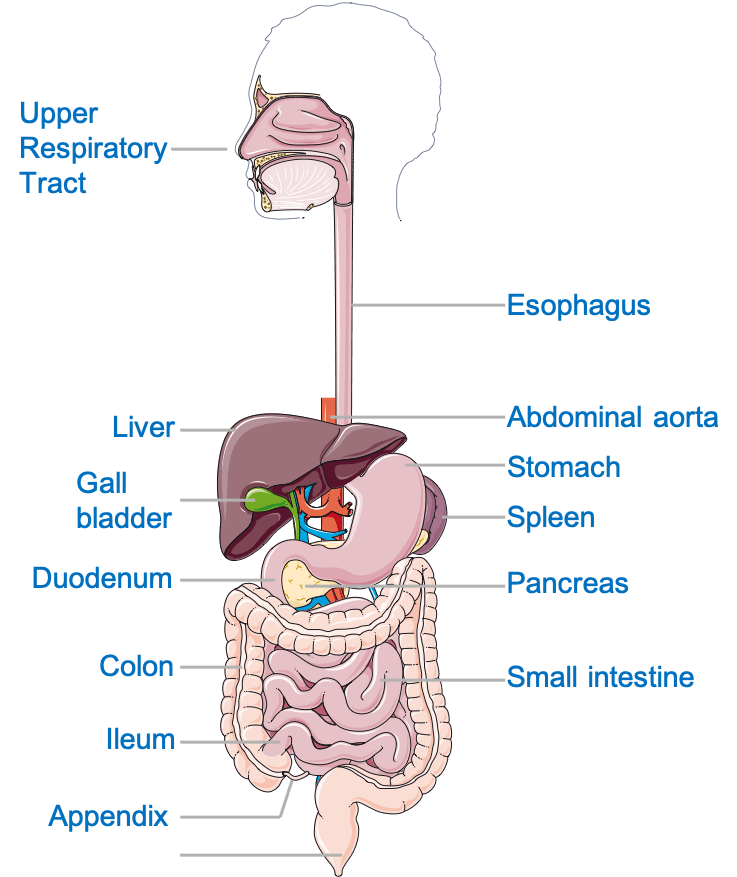
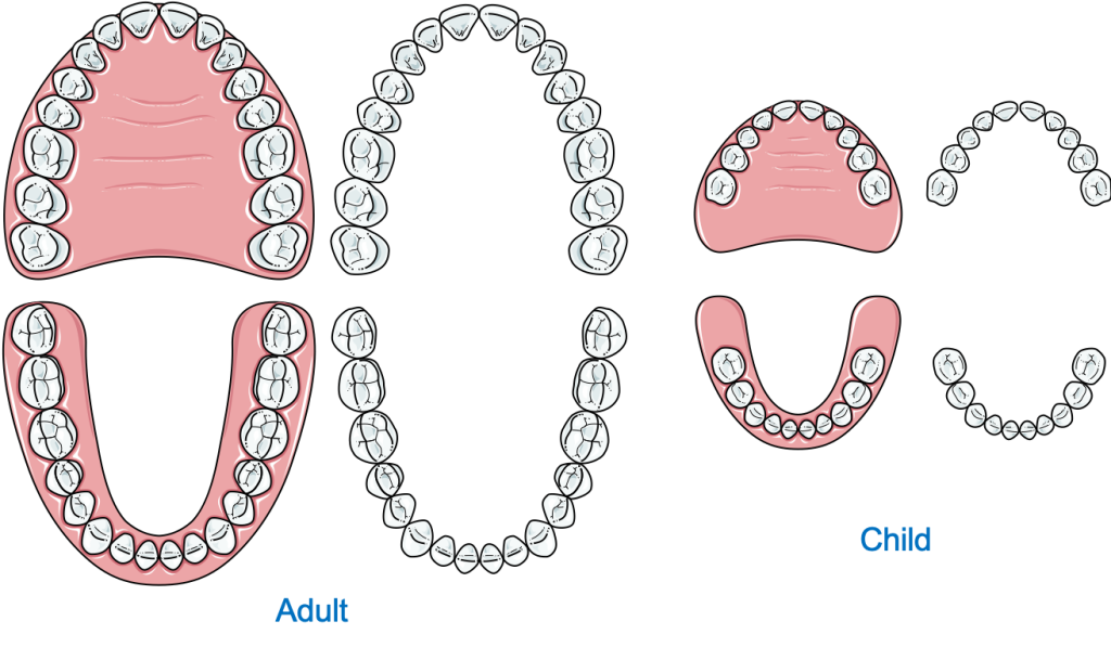
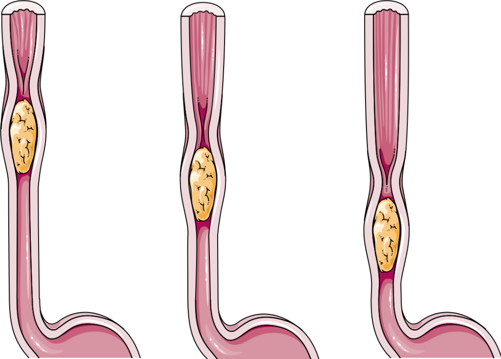
Mulut adalah titik awal dari saluran GI. Sejumlah besar pencernaan mekanis (pemecahan fisik makanan menjadi potongan-potongan kecil) terjadi di mulut. Mulut juga membantu melumasi makanan dengan air liur yang membantunya bergerak di sepanjang faring dan kerongkongan. Omong-omong, kerongkongan adalah tabung berotot yang menghubungkan mulut dengan perut dan menyediakan jalan bagi bolus (bola makanan setengah padat yang telah dikunyah dan dicampur dengan air liur).
Makanan yang mencapai lambung disimpan dan dicerna, baik secara mekanis maupun kimiawi. Pencernaan kimiawi mengacu pada proses di mana tubuh memecah molekul makanan kompleks yang tidak larut seperti pati menjadi molekul larut yang lebih kecil yaitu glukosa, asam amino, dan asam lemak. Itu perut adalah kantong berotot kuat yang berkontraksi secara berkala, memecah dan mencampur makanan dengan sekresi lambung; campuran air, asam klorida, dan protease (enzim pencerna protein). Ini memiliki kelengkungan yang lebih kecil dan lebih besar bersama dengan antrum dan pilorus. Kimus, makanan setengah cerna yang bercampur dengan sekresi lambung, didorong oleh peristaltik (gerakan seperti gelombang) ke dalam usus di mana sisa makanan akan dicerna dan diserap.
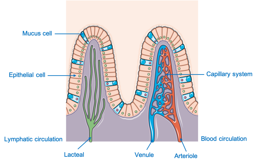
Usus halus dimulai dari pilorus lambung dan dibagi menjadi tiga bagian; duodenum, jejunum, dan ileum. Sebagian besar pencernaan terjadi di duodenum dan jejunum awal. Ini adalah struktur luminal sepanjang 5 meter dengan epitel khusus — batas sikat.
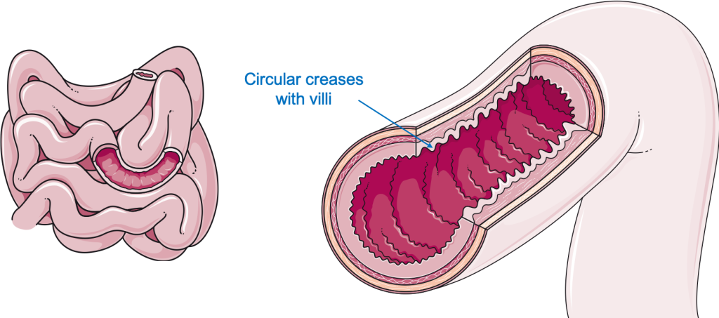
Epitel ini mengandung banyak vili dan mikrovili di seluruh permukaannya. Vili dan mikrovili adalah proyeksi kecil seperti jari yang sangat meningkatkan luas permukaan (area yang tersedia untuk penyerapan). Mikrovili ini juga membuat usus tampak seperti handuk. Setelah sebagian besar nutrisi yang larut yaitu asam amino, glukosa, fruktosa, asam lemak gliserol diserap, makanan didorong menuju anus di usus besar.
Usus besar yaitu, usus besar dimulai dari persimpangan ileocecal dan meluas ke rektum. Ascending, transversal, descending, dan kolon sigmoid adalah bagian dari usus besar. Fungsi utama usus besar adalah menyerap air dan elektrolit dari sisa makanan yang tidak tercerna. Usus besar juga memiliki serangkaian ikatan pita otot yang disebut tinea-coli yang berkontraksi untuk menghasilkan gerakan massal di seluruh usus besar dan membantu mendorong tinja ke dalam rektum.
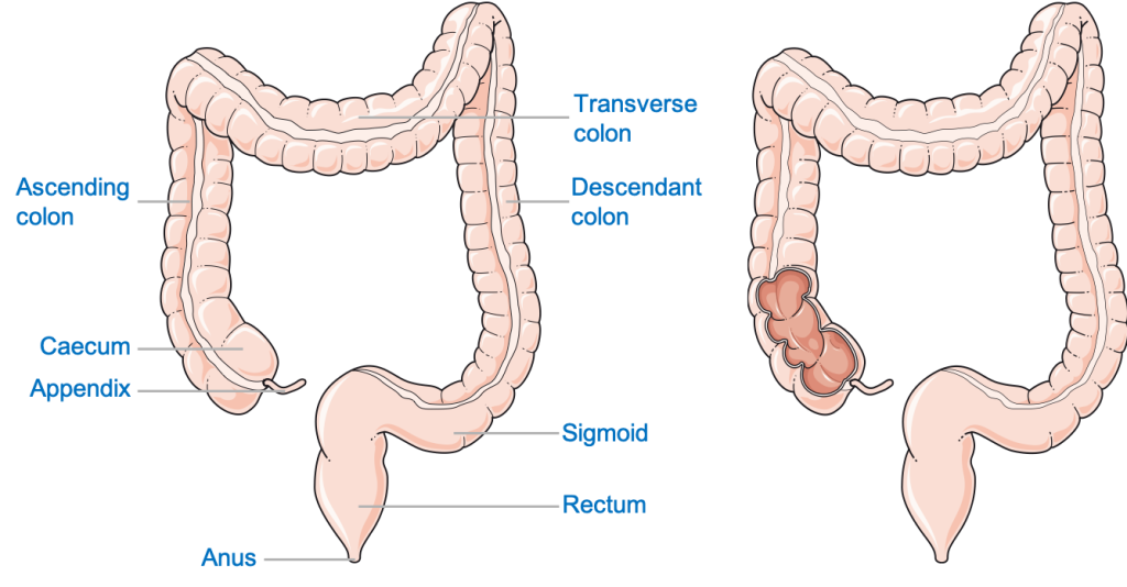
Rektum menghubungkan kolon sigmoid ke anus. Kotoran disimpan sementara di rektum hingga dikeluarkan dari tubuh melalui anus melalui proses yang disebut defekasi.
Hati memainkan fungsi vital dalam menjaga kesehatan tubuh. Pada kebanyakan orang, letaknya di daerah perut kanan atas, di bawah kubah diafragma kanan dan paru-paru kanan.
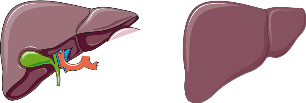
Terlepas dari banyak fungsi lainnya seperti detoksifikasi zat beracun eksogen, menyimpan nutrisi, dan sintesis protein dan trigliserida (lemak), hati juga memproduksi empedu. Empedu disimpan sementara di kantong empedu (melekat pada hati) dan disekresikan ke dalam usus kecil melalui saluran empedu. Empedu mengemulsi molekul lemak besar menjadi tetesan lemak yang lebih kecil yang dapat dicerna secara efektif oleh lipase (enzim pencerna lemak).
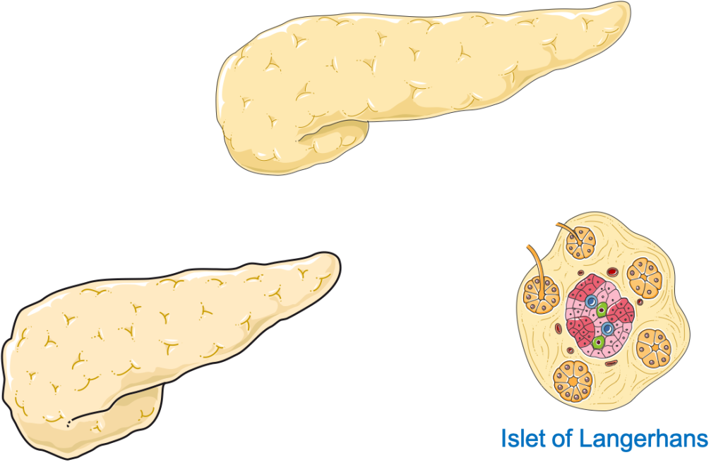
Pankreas adalah organ berbentuk daun dan berdasarkan fungsinya dibagi menjadi dua bagian: pankreas eksokrin dan endokrin (penghasil hormon). Pankreas eksokrin bertanggung jawab untuk memproduksi semua enzim pencernaan utama yang diperlukan untuk pemecahan makanan. Sekresi pankreas adalah campuran dari:
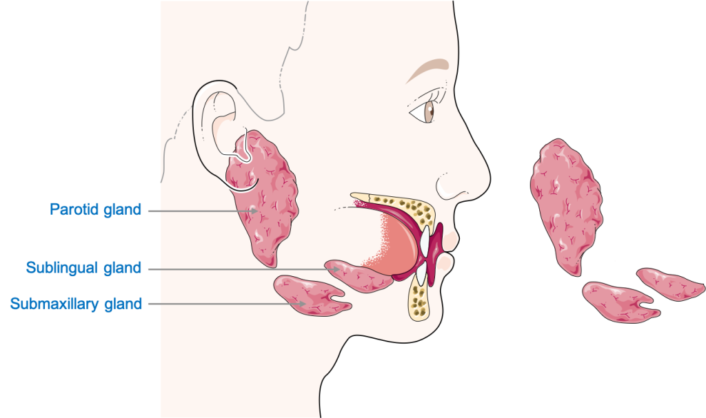
Saliva adalah sekresi encer, diproduksi oleh struktur kelenjar di rongga bukal, bercampur dengan enzim misalnya karbohidrat (enzim pencerna karbohidrat) dan lipase. Ini membantu melunakkan bolus makanan dan mencerna sebagian makanan, terutama pati (karbohidrat) yang ada dalam makanan. Ada tiga kelenjar ludah bilateral (ada di kedua sisi) yang ada di tubuh kita. Kelenjar parotis, kelenjar submandibular, dan kelenjar sublingual. Kelenjar parotid, hadir di bawah telinga, adalah kelenjar ludah utama. Seperti namanya, kelenjar submandibular terletak di bawah rahang bawah (tulang rahang) dan kelenjar sublingual di bawah lidah.
Pasokan neurovaskular mengacu pada suplai darah dan suplai saraf, koneksi yang sangat penting untuk menjaga organ tetap hidup. Pencernaan disuplai oleh satu set saraf dan pembuluh darah yang berbeda.
Rongga bukal dan struktur di dalamnya dipersarafi oleh saraf kranial (CN), saraf yang berasal langsung dari otak atau batang otak. Sebagian besar persarafan adalah oleh CN V (5), saraf trigeminal, bersama dengan CN IX (9) dan CN X (10). CN ke-10, saraf Vagus, memasok sebagian besar saluran GI. Stimulasi melalui CN X meningkatkan peristaltik dan sekresi di seluruh saluran GI. Fitur kunci lain yang perlu diperhatikan adalah bahwa persarafan sebagian besar sistem pencernaan berasal dari sistem saraf otonom, yaitu tidak berada di bawah kendali sukarela. Hati dan pankreas dipersarafi oleh saraf vagal dan splanchnic (simpatis).
Bagian bawah kanalis analis, di bawah garis pectinate, berasal dari saraf somatik (volunter), saraf pudendal. Ini memberi kita kendali atas buang air besar.
Pasokan vaskular mulut melibatkan cabang yang berbeda dari Arteri Karotis Eksternal (ECA) misalnya, arteri lingual ke lidah. Drainase vena mulut melalui serangkaian vena kecil yang akhirnya mengalir ke vena jugularis interna. Esofagus, lambung, dan bagian proksimal (atas) duodenum disuplai oleh cabang arteri Celiac (cabang aorta perut) dan dikeringkan oleh vena yang berdekatan kembali ke vena celiac. Bagian distal duodenum, jejunum, ileum, dan dua pertiga kolon transversal disuplai oleh Arteri Mesenterika Superior (cabang aorta perut). Sepertiga terakhir dari kolon transversal, kolon desenden dan sigmoid, dan saluran anus hingga garis pektinat disuplai oleh Arteri Mesenterika Inferior (cabang dari aorta perut). Di bawah garis pektinat, saluran anus disuplai oleh Arteri Pudendal. Drainase vena dari struktur ini adalah melalui vena dari arteri yang sesuai. Sebagian besar pankreas disuplai oleh cabang-cabang dari arteri Limpa (cabang Arteri Celiac) dan dikeringkan oleh vena lienalis.
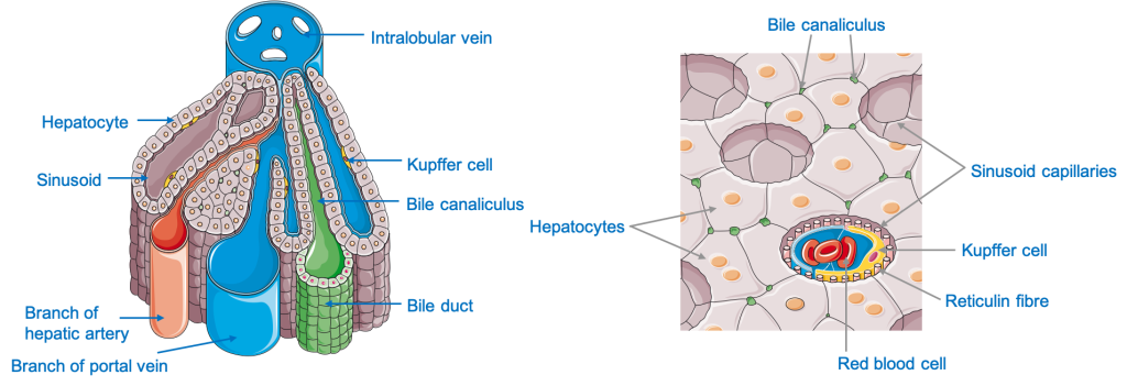
Hati memiliki kepentingan khusus karena terhubung ke saluran GI oleh vena Portal Hepatik yang memasok darah yang kaya nutrisi ke hati. Parenkim hati (jaringan) disuplai oleh arteri hepatika, yang berasal dari arteri Celiac, dan dialirkan oleh vena hepatik: cabang-cabang inferior vena cava.
Pelajari lebih lanjut tentang hubungan spasial 3D organ pencernaan dengan brilian ini model anatomi seukuran manusia.
Konten yang dibagikan di situs web Health Literacy Hub disediakan untuk tujuan informasi saja dan tidak dimaksudkan untuk menggantikan saran, diagnosis, atau perawatan yang ditawarkan oleh profesional medis yang memenuhi syarat di Negara Bagian atau Negara Anda. Pembaca didorong untuk mengkonfirmasi informasi yang diberikan dengan sumber lain dan untuk mencari nasihat dari praktisi medis yang memenuhi syarat dengan pertanyaan apapun yang mungkin mereka miliki mengenai kesehatan mereka. Health Literacy Hub tidak bertanggung jawab atas segala konsekuensi langsung atau tidak langsung yang timbul dari penerapan materi yang disediakan.


