
上腕骨–上腕
上腕骨に関するおもしろ情報
上腕骨は上腕/手足の長骨です。
上腕骨に関する3つの興味深い事実は次のとおりです。
1.上腕骨は、上肢で最も長い骨です。
2. Located between the shoulder and the elbow, the humerus is responsible for supporting the upper arm and performing daily movements such as eating and carrying objects.
3. The humerus is often referred to as the “funny bone’. However, the tingling sensation that comes from hitting your humerus is due to compression of the ulnar nerve that curves around the elbow.
この記事では、この骨の構造と機能を詳細に学び、上腕骨骨折の結果について説明します。
構造
The humerus consists of three parts; a humeral shaft, upper end, and lower end. The upper end of the bone is round to make the head. On the other hand, the lower end of the humerus is expanded and flattened from side to side.
Anatomical parts of the humerus consist of the head, anatomical/surgical neck, shaft, epicondyles (upper and lower ends), tubercles (greater and lesser for attachment of muscles of the arm), and capitulum (for articulation at the elbow).
Head and upper end
The Head of the humerus is rounded. In the upper end of the bone, there are other structures as the greater tubercle, lesser tubercle, bicipital groove, or intertubercle groove for muscular attachments.
Shaft
The shaft of the humerus is rounded in its upper half and triangular in the lower half. The shaft of the humerus consists of surfaces and borders for muscle and tendon attachments.
Lower end
The lower end of the humerus forms structures known as condyles that expand from one side to the other. The lower end contains a few non-articulating and articulating structures (for forming a joint with the elbow)
上腕骨の境界
To understand the borders of the humerus, you need to understand the definition of a few basic anatomical terms.
- Anterior (or ventral): describes the front or direction towards the front of the body.
- Posterior (or dorsal): this describes the back or direction toward the back of the body.
- Superior (or cranial): a position above or higher than another part of the body.
- Inferior (or caudal): a position below or lower than another part of the body.
- Lateral: describes the side or direction toward the side of the body. Example: The thumb is farther away from the body than the other digits, so it is lateral to the body
- Medial: describes the middle or direction toward the middle of the body. Example: The big toe is closer to the body than the other digits, so it is medial to the body.
- Proximal describes a position in a limb that is nearer to a certain point of attachment on the limb e.g. the elbow.
- Distal describes a position in a limb that is farther from a certain point of attachment.
- Superficial describes a position closer to the surface of the body. E.g. The skin is superficial to the bones.
- Deep describes a position farther from the surface of the body. E.g The brain is deep to the skull.
内側の境界線
- Its upper part of the medial border forms a groove for muscular attachment.
- The middle portion of the medial border is rough stripped.
- The medial border continues with the medial supracondylar ridge on the lower end of the humerus.
前縁
- The upper part of the lateral border forms a groove.
- The middle portion forms a projection of bone known as deltoid tuberosity.
- The lower end of the anterior border is round-shaped and smooth.
横方向の境界
- The upper end of the lateral border is not prominent or noticeable.
- The middle portion of the lateral border contains a spinal and radial groove for muscular attachment.
- The lower end of the lateral border forms a lateral supracondylar ridge.
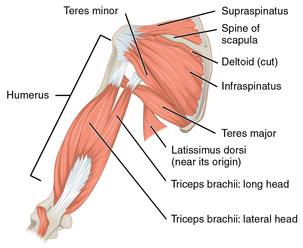
筋肉のアタッチメント
Several muscles of the arm, 肘, shoulder, and forearm attach, start or end on the humerus bone, they include :
- Supraspinatus
- Infraspinatus
- Pectoralis major
- Teres major
- Latissimus dorsi
- Deltoid
- Tricep brachii etc
神経供給
The 腋窩 からの神経 腕神経叢 wraps around the neck of the humerus. It provides innervation to teres minor, rotator cuff muscles, and deltoid. The radial nerve, which is also one of the five branches of the brachial plexus innervates multiple regions of the forearm, including the heads of the triceps brachii muscle of the upper arm.
血液供給
For the upper end of the humerus, the primary blood supply is from posterior and anterior circumflex humeral arteries which are branches of the axillary artery. Profunda brachii artery gives blood supply to the rest of the humerus.
上腕骨の機能
The two integral functions of the humerus in the body are support and movement.
The humerus forms a joint with the shoulder or 肩甲骨 and an 肘 とのジョイント 尺骨 and radius bones. Several movements occur at the shoulder joint, however just two movements; extension and flexion are possible on the 肘 ジョイント。
上腕骨とその関節で考えられる動きは次のとおりです。
- 循環–肩関節の回転
- 内転–腕の体への動き
- 誘拐–体から離れる腕の動き
- Extension of the arm – the movement of the arm behind the torso
- Flexion of the arm – the movement of the arm in front of the torso or overhead
- の拡張 肘 –腕を全開または伸ばす
- の屈曲 肘 –前腕の体への動き
臨床的関連性および関連疾患
上腕骨骨折
The most common pathology or injury of humerus bone is a fracture. The most common fracture sites for humerus are the:
- Humeral neck
- 上腕骨のシャフト
- 上腕骨顆上領域
上腕骨頭の骨折は、上腕骨頭の血液供給が不十分なため、治癒が遅い。
The most common fracture at a young age is a supracondylar fracture. The cause of this fracture is a fall or injury on the outstretched hand. In this fracture, the lower end of the bone tends to displace backward. Thus, it makes the 肘 prominent. In this fracture, the most commonly injured nerve is the median nerve.
Dislocations
A dislocation is an injury in which the ends of your bones are forced out from their normal positions in your joints. The common causes include trauma resulting from a fall, an auto accident, or a collision during contact or high-speed sports.
Dislocations are common problems with the shoulder joint. They include:
- 前方脱臼
- 後部脱臼
- 劣った脱臼
The most common dislocation for the humerus head is inferior dislocation due to a lack of structural support in the articulating surfaces of the shoulder joint.
- Gallinet D、Clappaz P、Garbuio P、Tropet Y、Obert L. 3つまたは4つの部分が複雑な近位上腕骨骨折:半関節形成術と逆プロテーゼ:40例の比較研究。整形外科および外傷学:手術および研究。 2009年2月1日; 95(1):48-55。
- Asharani SK、NingaiahA。上腕骨の栄養孔に関する研究。 Int J AnatRes。 2016; 4(3):2706-9。
- OrtnerDJ。上腕骨の遠位関節面における変性骨変化の説明と分類。 American Journal of PhysicalAnthropology。 1968年3月; 28(2):139-55。
- Zlotolow DA、Catalano III LW、Barron AO、GlickelSZ。上腕骨の外科的露出。 JAAOS-米国整形外科学会誌。 2006年12月1日; 14(13):754-65。
- Guse TR、OstrumRF。上腕骨周囲の橈骨神経の外科的解剖学。臨床整形外科および関連する研究。 1995年11月1日(320):149-53。
- DeLude JA、Bicknell RT、MacKenzie GA、Ferreira LM、Dunning CE、King GJ、Johnson JA、Drosdowech DS上腕骨の両側の解剖学の人体測定研究。肩と肘の手術のジャーナル。 2007年7月1日; 16(4):477-83。
- ラフCB、ジョーンズHH。上腕骨と脛骨の皮質骨における両側非対称性—性別と年齢の要因。人間生物学。 1981年2月1日:69-86。
- ファーンズワースCL、シルバPD、ムバラクSJ上腕骨顆上骨折の病因。 Journal of PediatricOrthopaedics。 1998年1月1日; 18(1):38-42。
- タナーMW、コフィールドRH。上腕骨近位部の骨折および骨折脱臼のための人工関節形成術。臨床整形外科および関連する研究。 1983年10月1日(179):116-28。
Health Literacy Hub Webサイトで共有されるコンテンツは、情報提供のみを目的として提供されており、州または国の資格のある医療専門家が提供するアドバイス、診断、または治療に代わるものではありません。読者は、他の情報源から提供された情報を確認し、健康に関して質問がある場合は資格のある開業医のアドバイスを求めることをお勧めします。 Health Literacy Hubは、提供された資料の適用から生じる直接的または間接的な結果に対して責任を負いません。


