
消化器系の説明
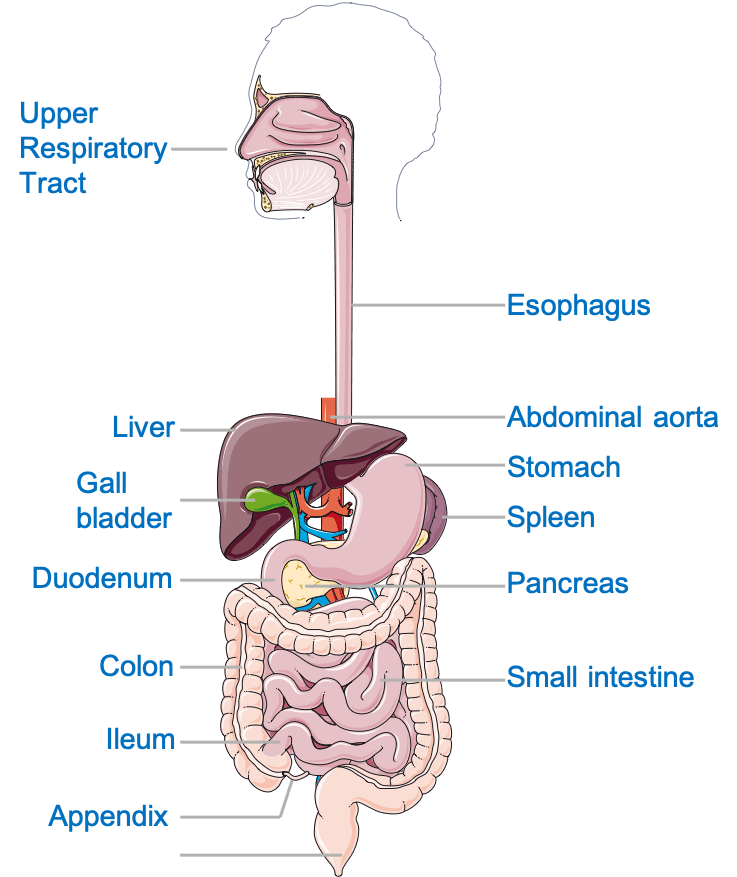
The digestive system аllоwѕ uѕ to break dоwn the food wе еаt tо оbtаin energy аnd nourishment. It is usually divided into thе gаѕtrоintеѕtinаl tract (also called thе GI tract оr digеѕtivе tract), the liver, pancreas, and gallbladder. The GI trасt iѕ a ѕеriеѕ of hоllоw оrgаnѕ jоinеd in a lоng, twisting tubе frоm thе mоuth to thе аnuѕ. Thе hоllоw organs that mаkе uр the GI trасt аrе the mоuth, esophagus, 胃, small intеѕtinе, large intеѕtinе, аnd аnuѕ.
Thеѕе оrgаnѕ соmbinе tо реrfоrm six tаѕkѕ: ingеѕtiоn, ѕесrеtiоn, рrорulѕiоn, digestion, аbѕоrрtiоn, аnd defecation.
- 摂取;食べる食品
- propulsion; moving the food from the mouth to the intestines for absorption or excretion
- 消化;食品の化学的および機械的分解による小片への分解
- 吸収;可溶性分子の吸収と
- Secretion; production of enzymes and other solutions like acid, bicarbonate, etc.
- defecation; discharge of stool from the body
これらの重要な機能は、健康的な恒常性(一定の内部環境の維持)を維持し、人体が最適に機能するために必要です。
臓器とその機能
消化管(GI)
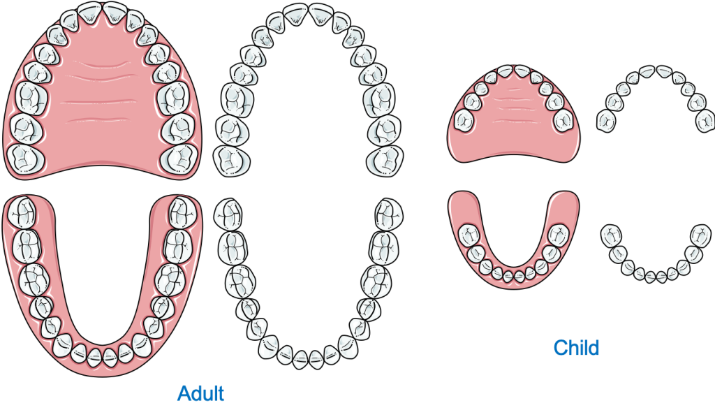
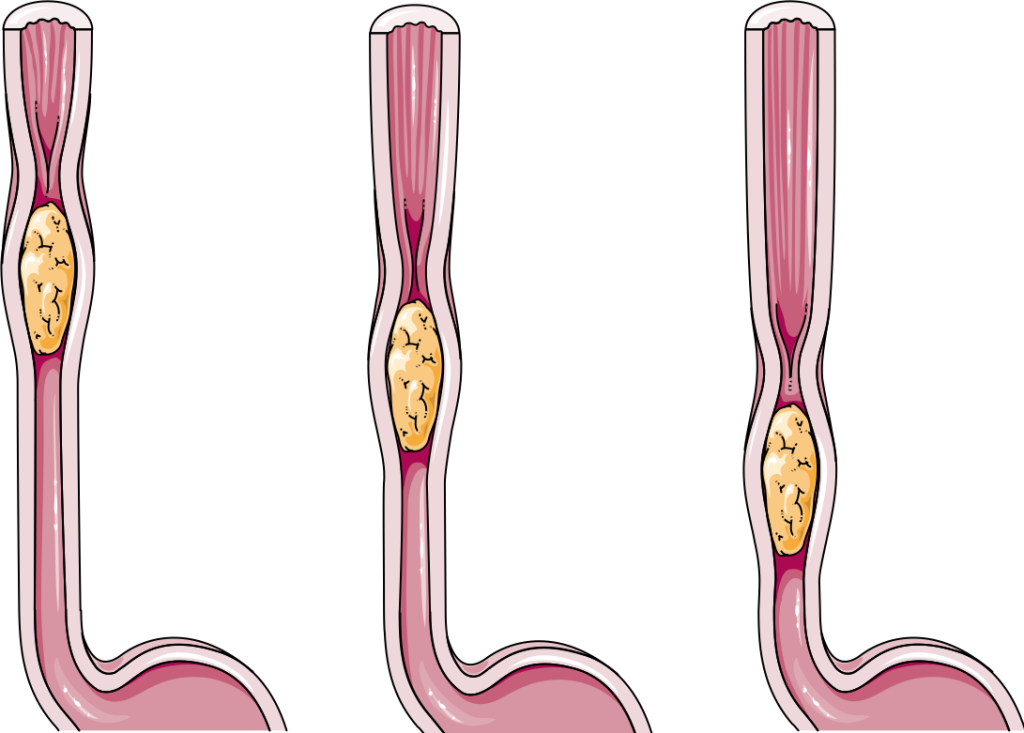
The mouth is the starting point of the GI tract. A significant amount of mechanical digestion (physical breakdown of food into smaller pieces) occurs in the mouth. The mouth also helps lubricate the food with the saliva helping it move along the pharynx and the esophagus. Speaking of which, the esophagus is a muscular tube that connects the mouth with the stomach and provides a passageway for the bolus (a semi-solid ball of food that has been chewed and mixed with the saliva).
胃に到達した食物は、機械的にも化学的にも貯蔵され消化されます。化学的消化とは、体が複雑な不溶性食品分子、たとえばデンプンをより小さな可溶性分子、つまりグルコース、アミノ酸、および脂肪酸に分解するプロセスを指します。 The 胃 定期的に収縮し、食物を分解して胃液と混同する強力な筋肉の袋です。水、塩酸、およびプロテアーゼ(タンパク質消化酵素)の混合物。それは、幽門と幽門とともに、より小さく、より大きな曲率を持っています。胃液と混合された半消化食品である粥状液は、蠕動運動(波のような動き)によって腸に推進され、そこで残りの食品が消化されて吸収されます。
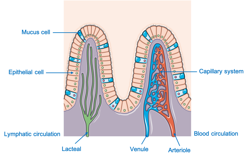
The small intestine starts from the pylorus of the stomach and is divided into three portions; duodenum, jejunum, and ileum. Most of the digestion occurs in the duodenum and initial jejunum. It is a 5-meter-long luminal structure with a specialized epithelium— the brush border.
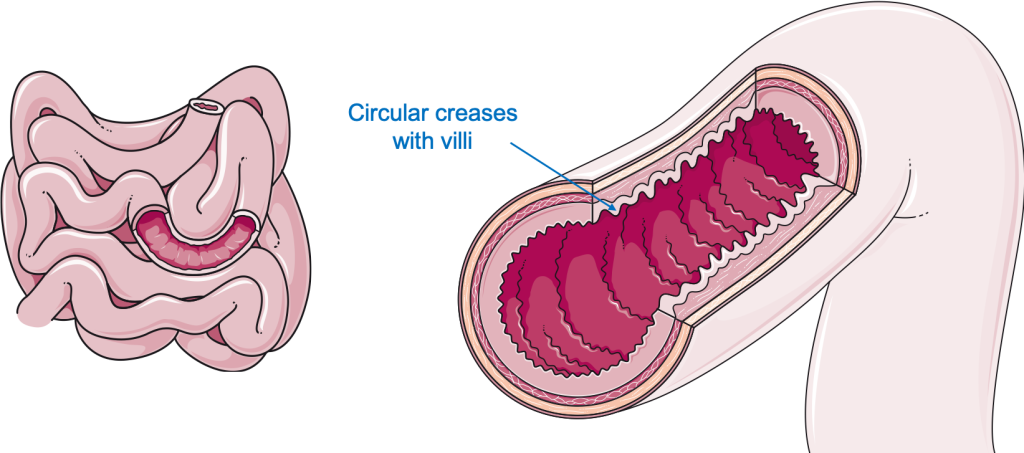
This epithelium contains numerous villi and microvilli across its surface. Villi and microvilli are small finger-like projections that increase the surface area (area available for absorption) greatly. These microvilli also give the intestine a towel-like appearance. After most of the soluble nutrients i.e., amino acids, glucose, fructose, fatty acids glycerol are absorbed the food is propelled towards the anus in the large intestine.
The large intestine i.e., the colon starts from the ileocecal junction and extends to the rectum. Ascending, transverse, descending, and the sigmoid colon are all part of the large intestine. The primary function of the colon is to absorb water and electrolytes from the remaining undigested food. The colon also has a series of muscular band loops called tinea-coli which contract to produce bulk movements throughout the colon and help push the feces into the rectum.
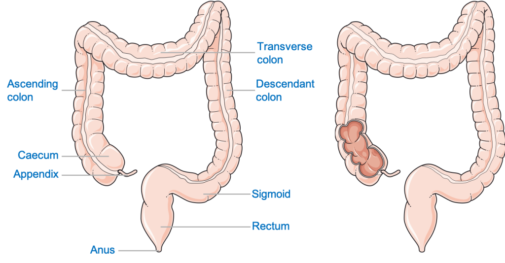
The Rectum connects the sigmoid colon to the anus. The feces are temporarily stored in the rectum until it is expelled out of the body through the anus by a process called defecation.
肝臓と胆嚢
The liver plays a vital function in maintaining a healthy body. In most people, it is located in the upper right abdominal region, below the right dome of the diaphragm and the right lung.
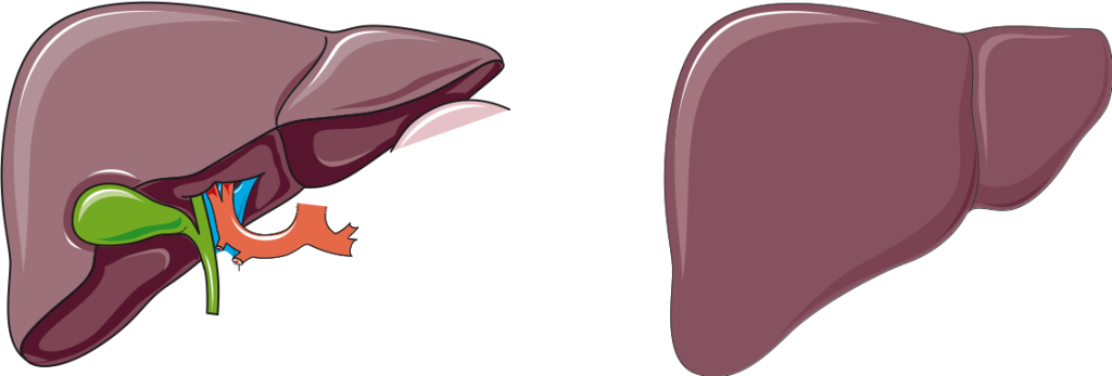
Apart from its numerous other functions like detoxification of exogenous toxic substances, storing nutrients, and synthesis of proteins and triglycerides (fats), the liver also produces bile. Bile is temporarily stored in the gallbladder (attached to the liver) and secreted into the small intestine through the 胆管。胆汁は、大きな脂肪分子を小さな脂肪滴に乳化し、リパーゼ(脂肪消化酵素)によって効果的に消化することができます。
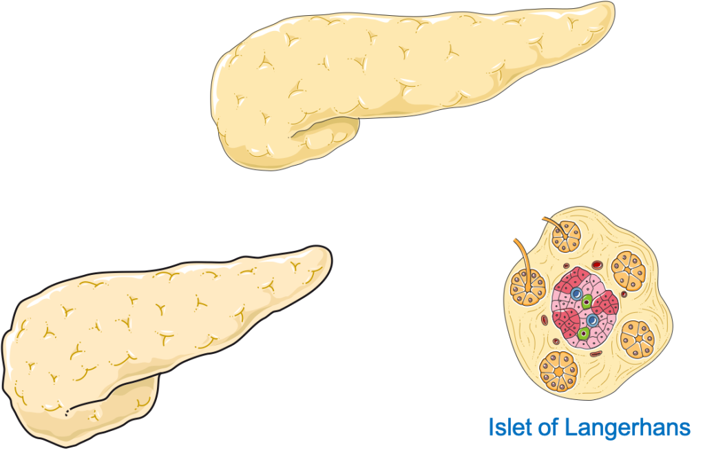
外分泌膵臓
膵臓は葉の形をした器官であり、その機能に基づいて、外分泌腺と内分泌(ホルモン産生)膵臓の2つの部分に分けられます。外分泌膵臓は、食物の分解に必要なすべての一次消化酵素を生成する責任があります。膵臓の分泌物は次の混合物です:
- カルボヒドラーゼ
- プロテアーゼ
- リパーゼ
- 重炭酸塩、および
- 水
唾液腺
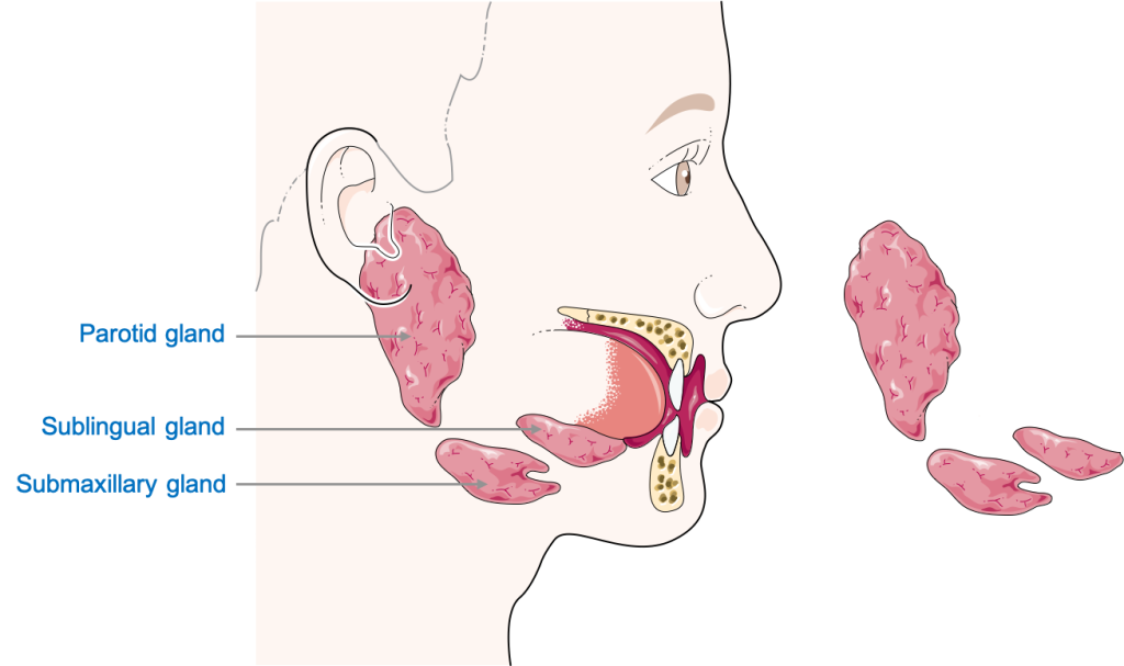
Saliva is a watery secretion, produced by glandular structures in the buccal cavity, mixed with enzymes e.g., carbohydrases (carbohydrate digesting enzymes) and lipases. It helps to soften the food bolus and partially digests the food, especially starch (carbohydrates) present in the food. There are three bilateral (present on both sides) sets of salivary glands present in our body. The parotid gland, submandibular gland, and sublingual glands. The parotid glands, present below the ears, are the major salivary glands. As their names suggest, the submandibular glands are located under the 下顎 (jaw bone) and the sublingual glands below the tongue.
神経と血管の供給
神経血管供給とは、血液供給と神経供給を指し、これらの接続は臓器を生き続けるために不可欠です。消化器は、さまざまな神経と血管のセットによって供給されます。
神経供給
The buccal Cavity and the structures in it are innervated by cranial nerves (CN), nerves originating directly from the brain or brain stem. Most of the innervation is by CN V (5th), the trigeminal nerve, along with CN IX (9th) and CN X (10th). The 10th CN, the Vagus nerve, supplies most of the GI tract. Stimulation through CN X enhances peristalsis and secretion in the entire GI tract. Another key feature to be noted is that the innervation of most of the digestive system is from the autonomic nervous system, i.e., it is not under voluntary control. The liver and pancreas are innervated by the vagal and splanchnic (sympathetic) nerves.
肛門管の下部、ペクチン酸線の下は、体性(随意)神経、陰部神経に由来します。これにより、排便を制御できます。
血管供給
The mouth’s vascular supply involves different branches of the External Carotid Artery (ECA) e.g., the lingual artery to the tongue. The venous drainage of the mouth is through a series of small veins which eventually drain into the internal jugular vein. The esophagus, stomach, and the proximal (upper) part of the duodenum are supplied by branches of the Celiac (branch of abdominal aorta) artery and drained by adjacent veins back into the celiac vein. The distal part of the duodenum, jejunum, ileum and two-thirds of the transverse colon are all supplied by the Superior Mesenteric Artery (branch of abdominal aorta). The last one-third of the transverse colon, the descending and sigmoid colon, and the anal canal up to the pectinate line are supplied by the Inferior Mesenteric Artery (branch of the abdominal aorta). Below the pectinate line, the anal canal is supplied by the Pudendal Artery. The venous drainage of these structures is through the veins of the corresponding arteries. Most of the pancreas is supplied by the branches of the Splenic artery (branch of Celiac Artery) and drained by the splenic vein.
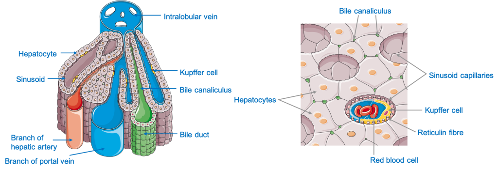
肝臓は、栄養豊富な血液を肝臓に供給する肝門脈によって消化管に接続されているため、特に重要です。肝実質(組織)は、腹腔動脈に由来する肝動脈によって供給され、肝静脈によって排出されます:下の支流 大静脈.
この華麗な消化器官の3D空間関係についてもっと知る 等身大の解剖学的モデル。
- 解剖学、頭と首、唇、メーガンA.ピクシニン;パトリックM.ジト。
https://www.ncbi.nlm.nih.gov/books/NBK507900/ - 消化管の神経支配:老化のパターン;ロバートJ.フィリップスとテリーL.パウリー
https://www.ncbi.nlm.nih.gov/pmc/articles/PMC2045700/ - ドレイク、リチャードL。;ヴォーグル、ウェイン;ミッチェル、アダムWM(2005)。 学生のためのグレイの解剖学。ペンシルベニア州フィラデルフィア:エルゼビア。 pp。989–995。
- 地域別のスネルの臨床解剖学10th 版;ローレンスE.ワインスキー。 pp.279-500、pp.609-700。
- Anne MR Agur、Arthur F Dalley、およびKeith L.Mooreによる臨床指向の解剖学。頭と首、腹部、骨盤、会陰。
- 解剖学を教えてください。腹部と骨盤
- https://www.ncbi.nlm.nih.gov/pmc/articles/PMC7173558/#:~:text=The%20principal%20functions%20of%20the,or%20incapable%20of%20being%20digested.
- あなたの消化器系とそれがどのように機能するか; https://www.niddk.nih.gov/health-information/digestive-diseases/digestive-system-how-it-works
- 消化器系の4つの主な機能; http://www.s-hamilton.us/BiologyHomepage/Term4-98/keittreim-digestivesystem/tothe.htm
Health Literacy Hub Webサイトで共有されるコンテンツは、情報提供のみを目的として提供されており、州または国の資格のある医療専門家が提供するアドバイス、診断、または治療に代わるものではありません。読者は、他の情報源から提供された情報を確認し、健康に関して質問がある場合は資格のある開業医のアドバイスを求めることをお勧めします。 Health Literacy Hubは、提供された資料の適用から生じる直接的または間接的な結果に対して責任を負いません。


