Modelo Anatômico 3D
Adicione outra dimensão ao seu aprendizado com modelos anatômicos educacionais masculinos e femininos totalmente interativos.
Aprender sobre a anatomia humana nunca foi tão divertido!
Comprar
O nervo óptico é o feixe de cerca de cem milhões de fibras nervosas que transportam informações da retina para o cérebro. O nervo óptico é o que permite que você veja, e sem ele, a maioria das pessoas com deficiência visual são cegas. Somos fatos interessantes para além disso, é que ele envia muito mais em cerca de 12 milissegundos, 301ttp2t de inocente, por exemplo, você pode vìvula, e você pode sequer se alguém está desse tipo, que eles piscarem mais.
Neste artigo, veremos a anatomia do nervo óptico – seu curso, funções sensoriais e relevância clínica.
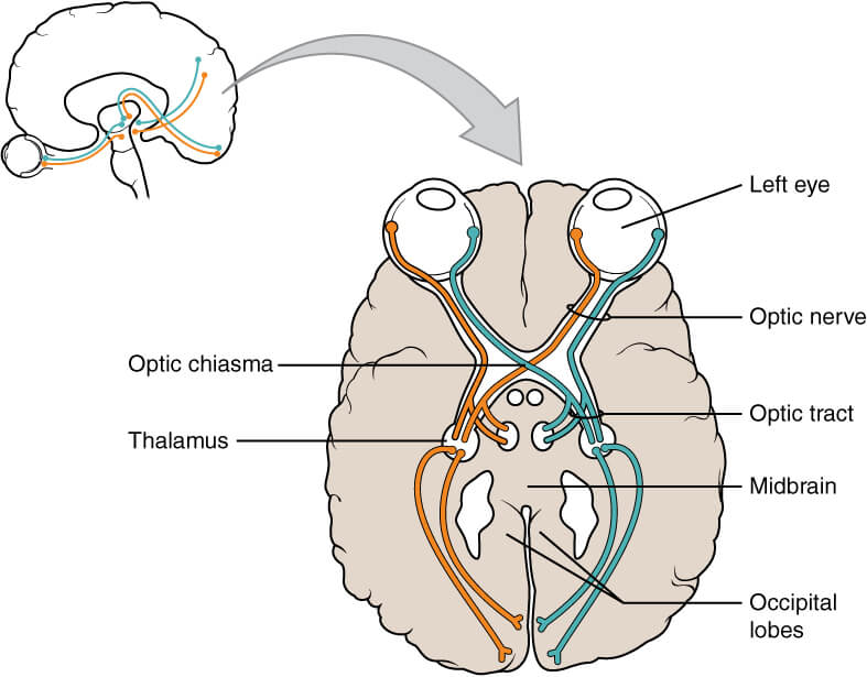
A via anatômica do nervo óptico descreve a transmissão de informações sensoriais especiais da retina do olho para o córtex visual primário do cérebro. Pode ser dividido em componentes extracranianos (fora da cavidade craniana) e intracranianos (dentro da cavidade craniana).
Extracraniano
O nervo óptico é formado pela convergência da porção de células nervosas que transporta os impulsos nervosos para longe do corpo celular chamados axônios. Esses axônios vêm das células ganglionares da retina. Essas células, por sua vez, recebem impulsos dos receptores de luz do olho (os bastonetes e cones).
Após a formação do nervo óptico, o nervo deixa a órbita óssea através de uma passagem no osso esfenóide conhecido como canal óptico. Ele entra na cavidade craniana, correndo ao longo da superfície da parte central da base do crânio (a fossa craniana intermediária) próximo à glândula pituitária.
Intracraniano (A Via Visual)
Dentro da parte central da base do crânio, os nervos ópticos de cada olho se unem para formar o cisma óptico (um espaço em forma de X, diretamente na frente do hipotálamo). No quiasma, as fibras desse nervo da metade nasal de cada retina cruzam para o trato óptico oposto, enquanto as fibras das metades temporais permanecem no mesmo trato.
Seu campo visual refere-se à área total na qual os objetos ao lado podem ser vistos por seu olho enquanto você foca seus olhos em um ponto central.
Os axônios do LGN transportam informações visuais por meio de um caminho conhecido como radiação óptica. O caminho em si pode ser dividido em:
Uma vez no córtex visual, o cérebro processa os dados sensoriais e responde adequadamente.

O nervo óptico recebe uma ramificação direta da artéria oftálmica e da artéria central da retina. Também recebe ramificações das artérias coróides.
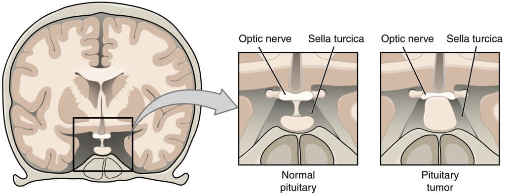
Adenoma Hipofisário
Um adenoma pituitário é um tumor da glândula pituitária. Dentro da parte central da base do crânio, a glândula pituitária fica próxima ao quiasma óptico. O aumento da glândula pituitária pode, portanto, afetar o funcionamento do nervo óptico.
A compressão do quiasma óptico afeta particularmente as fibras que cruzam a metade nasal de cada retina. Isso produz um defeito visual que afeta a visão periférica em ambos os olhos, conhecido como hemianopia bitemporal.
“Óculos de nervo óptico.” Nervo óptico, https://www.opticnerve.com/. Acessado em 3 de janeiro de 2022.
Distúrbios do Nervo Óptico. https://medlineplus.gov/opticnervedisorders.html. Acessado em 1 de janeiro de 2022.
"Nervo óptico." Kenhub, https://www.kenhub.com/en/library/anatomy/the-optic-nerve. Acessado em 3 de janeiro de 2022.
O conteúdo compartilhado no site Health Literacy Hub é fornecido apenas para fins informativos e não se destina a substituir conselhos, diagnósticos ou tratamentos oferecidos por profissionais médicos qualificados em seu Estado ou País. Os leitores são encorajados a confirmar as informações fornecidas com outras fontes e a procurar o conselho de um médico qualificado com qualquer dúvida que possam ter em relação à sua saúde. O Health Literacy Hub não se responsabiliza por qualquer consequência direta ou indireta decorrente da aplicação do material disponibilizado.