3D Anatomy Model
Add another dimension to your learning with fully-interactive educational male and female anatomical models.
Learning about the human anatomy has never been more fun!
Purchase
The humerus is the long bone of the upper arm/limb.
Here are three interesting facts about the humerus bone:
1. The humerus is the longest bone in the upper limb.
2. Located between the shoulder and the elbow, the humerus is responsible for supporting the upper arm and performing daily movements such as eating and carrying objects.
3. The humerus is often referred to as the “funny bone’. However, the tingling sensation that comes from hitting your humerus is due to compression of the ulnar nerve that curves around the elbow.
In this article, we will learn in detail the structure and function of this bone, and discuss the consequences of a humerus fracture.
The humerus consists of three parts; a humeral shaft, upper end, and lower end. The upper end of the bone is round to make the head. On the other hand, the lower end of the humerus is expanded and flattened from side to side.
Anatomical parts of the humerus consist of the head, anatomical/surgical neck, shaft, epicondyles (upper and lower ends), tubercles (greater and lesser for attachment of muscles of the arm), and capitulum (for articulation at the elbow).
The Head of the humerus is rounded. In the upper end of the bone, there are other structures as the greater tubercle, lesser tubercle, bicipital groove, or intertubercle groove for muscular attachments.
The shaft of the humerus is rounded in its upper half and triangular in the lower half. The shaft of the humerus consists of surfaces and borders for muscle and tendon attachments.
The lower end of the humerus forms structures known as condyles that expand from one side to the other. The lower end contains a few non-articulating and articulating structures (for forming a joint with the elbow)
To understand the borders of the humerus, you need to understand the definition of a few basic anatomical terms.
Several muscles of the arm, elbow, shoulder, and forearm attach, start or end on the humerus bone, they include :
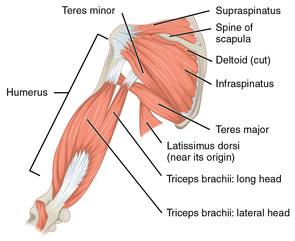
The axillary nerve from the brachial plexus wraps around the neck of the humerus. It provides innervation to teres minor, rotator cuff muscles, and deltoid. The radial nerve, which is also one of the five branches of the brachial plexus innervates multiple regions of the forearm, including the heads of the triceps brachii muscle of the upper arm.
For the upper end of the humerus, the primary blood supply is from posterior and anterior circumflex humeral arteries which are branches of the axillary artery. Profunda brachii artery gives blood supply to the rest of the humerus.
The two integral functions of the humerus in the body are support and movement.
The humerus forms a joint with the shoulder or scapula and an elbow joint with the ulna and radius bones. Several movements occur at the shoulder joint, however just two movements; extension and flexion are possible on the elbow joint.
Possible movements with the humerus and its joints are:
The most common pathology or injury of humerus bone is a fracture. The most common fracture sites for humerus are the:
Fractures at the humeral head are slow to heal due to the insufficient blood supply of the humeral head.
The most common fracture at a young age is a supracondylar fracture. The cause of this fracture is a fall or injury on the outstretched hand. In this fracture, the lower end of the bone tends to displace backward. Thus, it makes the elbow prominent. In this fracture, the most commonly injured nerve is the median nerve.
A dislocation is an injury in which the ends of your bones are forced out from their normal positions in your joints. The common causes include trauma resulting from a fall, an auto accident, or a collision during contact or high-speed sports.
Dislocations are common problems with the shoulder joint. They include:
The most common dislocation for the humerus head is inferior dislocation due to a lack of structural support in the articulating surfaces of the shoulder joint.
The content shared on the Health Literacy Hub website is provided for informational purposes only and it is not intended to replace advice, diagnosis, or treatment offered by qualified medical professionals in your State or Country. Readers are encouraged to confirm the information provided with other sources and to seek the advice of a qualified medical practitioner with any question they may have regarding their health. The Health Literacy Hub is not liable for any direct or indirect consequence arising from the application of the material provided.


