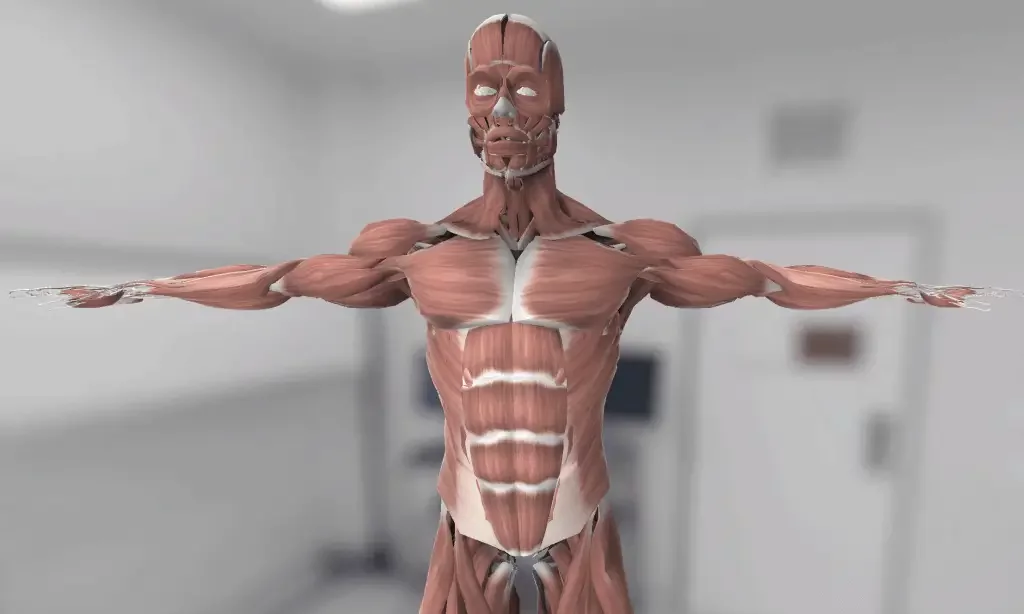
3D Anatomy Model
Add another dimension to your learning with fully-interactive educational male and female anatomical models.
Learning about the human anatomy has never been more fun!
Purchase
The excretory system as the name suggests is responsible to clear out waste from the body. Each and every cell in the body undergoes a million if not a billion chemical reactions per day. Each of these reactions results in many chemical compounds, some of which are metabolic waste.
Apart from cellular metabolism, the food we eat, and the medicine we take all produce toxic waste products that have to be removed from the body.
The essential functions of eliminating toxic waste, maintaining electrolyte balance, and maintaining fluid balance are performed by the excretory system of the body.
The excretory system is mainly composed of the kidneys, ureters, bladder, and urethra.
The primary function of the urinary system is to get rid of waste products in the body. Waste products including ammonia, urea, uric acid, etc are filtered by the kidneys and excreted in the urine. Drugs and other toxins are also excreted through the excretory system.
The kidneys and skin are responsible for maintaining osmotic and electrolyte balance in the body. The amount of urine and sweat is altered according to the needs of the body.
The excretory system consists of the urinary system and contributions from the liver, skin, and lungs.
Urinary System includes kidneys, ureters, urinary bladder, and urethra. The structure and function of these individual components of the urinary system are in the following section.
The main component of the urinary system is the kidney. Kidneys are bean-shaped organs located in the posterior part of the abdomen on the sides of the vertebral column. The kidneys have three layers around them. The outermost tough layer of connective tissue is the renal fascia. Under the renal fascia, there is a peri-renal fat capsule. The third and innermost covering of the kidneys is a renal capsule. Each kidney has three main parts.
The renal cortex is the outer granular part of the kidneys. The renal cortex has a granular appearance due to the presence of nephrons. Nephrons are considered the functional unit of the kidney.
The renal medulla is the inner part of the kidneys. The renal medulla has renal pyramids and renal columns.
The renal pelvis is the exit of the kidneys. The urine from the nephrons drains into the renal pelvis. The ureters are the continuation of the renal pelvis.
As said above, nephrons are the functional units of kidneys. Each nephron consists of the following parts.
Bowman’s capsule is the central part of the nephron. It is a cup-shaped structure and receives blood through afferent arterioles. The afferent arterioles divide into a network of capillaries in the bowman’s capsule called the glomerulus. Next to the bowman’s capsule is a proximal convoluted tubule; it is a convoluted tube extending down from the bowman’s capsule.
The third part of the nephron is a loop of Henle. The first part is a straight descending tubule that is a continuation of PCT. Then, there is a loop formation. Finally, there is ascending limb that connects with the distal convoluted tubule. After the loop of Henle, there is a distal convoluted tubule. The DCT opens in the collection duct. The collecting ducts open in the renal pelvis.
Nephrons are considered the main functional unit of the kidney. Hence, the functions of the nephron are considered the functions of the kidney. The function of the bowman’s capsule or glomeruli is to filter the blood. The PCT absorbs most of the water, glucose, and amino acids from the filtrate.
The loop of Henle and collecting duct respond to hormones. Their primary function is to adjust the osmolality of the urine according to the body’s needs. The PCT is the part that responds to hormones and plays a vital role in adjusting the excretion and reabsorption of electrolytes from the filtrate.
The individual functions of different parts ultimately lead to urine formation. Hence, the kidney function is to excrete waste materials and maintain the osmotic and electrolyte balance of the body.
Ureters are thin muscular tubes that are a continuation of the renal pelvis. Their function is to carry the urine from the kidneys to the urinary bladder.
The bladder is a balloon or sac-like organ. The wall of the bladder is made up of smooth muscles. The function of the bladder is to store urine. The bladder also has the role of contracting and passing urine in the urethra on nervous stimulation.
The urethra is a thin tube. Females have a shorter urethra compared to males. The urethra arises from the urinary bladder. Its function is to carry urine outside on the contraction of the bladder. The nervous system stimulates the opening of the urethra.
Your kidneys receive 20 percent of blood pumped by the heart with each beat. The blood supply of the kidneys is from the renal arteries. The renal artery is a direct branch of the abdominal aorta that arises just distal to SMA. The renal artery divides into the anterior and posterior divisions at the hilum of the kidney. These anterior and posterior divisions further split into five segmental arteries.
The segmental arteries divide into interlobar arteries, which subsequently divide into arcuate arteries. The arcuate arteries give rise to an afferent arteriole; the afferent arteriole further divides into glomerular capillaries. The filtration of the blood takes place at glomerular capillaries. Glomerular capillaries again unite to form efferent arterioles. Efferent arterioles provide blood to the outer two-thirds of the kidneys through the peritubular network. The peritubular network finally drains into the venous system.
The venous drainage of kidneys occurs through right and left renal veins, directly opening into the inferior vena cava.
The nerve supply of the kidneys is from the renal plexus. The renal plexus is formed by the branches of celiac and aorticorenal ganglia. It also receives branches from the lower thoracic splanchnic nerves and the first lumbar splanchnic nerve.
The blood Supply of the Ureters is segmental. The renal arteries supply the upper part of the ureters. The middle part of the ureters is supplied by the common iliac arteries and gonadal arteries. The distal portion of the ureters has blood supply from the branches of the internal iliac artery.
The venous drainage of the ureters is also segmental. It is carried by the mirror veins of the above-described arteries.
The nerve supply of the ureters is derived from three plexus; renal plexus, testicular/ovarian plexus, and hypogastric plexus.
The blood supply of the bladder is mainly from the internal iliac arteries. The superior vesical artery, a branch of the internal iliac artery, is the major blood supply of the artery. In males, the additional blood to the bladder is supplied by the inferior vesical artery. Vaginal arteries replace the inferior vesical artery in females.
The respective mirror veins of the above-described arteries finally carry the venous drainage of the bladder into an internal iliac vein.
The nerve supply of the bladder has sympathetic and parasympathetic components. The sympathetic nerve supply of the bladder is from the superior and inferior hypogastric plexus. The parasympathetic nerve supply of the bladder is from the pelvic splanchnic nerves.
The blood supply of the male urethra
Prostatic Urethra: Inferior vesical artery
Membranous Urethra: Bulbourethral Artery
Penile Urethra: Branches of Internal pudendal artery
The blood supply of the female urethra is carried through internal pudendal arteries mainly. The vaginal arteries and inferior vesical branches of vaginal arteries also contribute.
The venous drainage of the urethra is also carried by the mirror veins of the above-described arteries.
The nerve supply of the male urethra is from the prostatic plexus. And the nerve supply of the female urethra is from the vesical plexus and pudendal nerve.
1: Schulze A. (2001). Comparative anatomy of excretory organs in vestimentiferan tube worms (Pogonophora, Obturata). Journal of morphology, 250(1), 1–11. https://doi.org/10.1002/jmor.1054
2: Davis, L. E., Schmidt-Nielsen, B., & Stolte, H. (1976). Anatomy and ultrastructure of the excretory system of the lizard, Sceloporus cyanogenys. Journal of morphology, 149(3), 279–326. https://doi.org/10.1002/jmor.1051490302
3: Richardson M. (2006). The urinary system. Part 1–introduction. Nursing times, 102(40), 26–27. https://pubmed.ncbi.nlm.nih.gov/17042339/
4: Heading C. (1987). Anatomy and physiology of the urinary system. Nursing, 3(22), 812–814. https://pubmed.ncbi.nlm.nih.gov/3696557/
5: El-Bermani A. W. (1978). Anatomy of the urinary tract. Clinical obstetrics and gynecology, 21(3), 819–830. https://doi.org/10.1097/00003081-197809000-00018
6: Gómez, F. A., Ballesteros, L. E., & Estupiñán, H. Y. (2017). Anatomical study of the renal excretory system in pigs. A review of its characteristics as compared to its human counterpart. Folia morphologica, 76(2), 262–268. https://doi.org/10.5603/FM.a2016.0065
7: Hickling, D. R., Sun, T. T., & Wu, X. R. (2015). Anatomy and Physiology of the Urinary Tract: Relation to Host Defense and Microbial Infection. Microbiology spectrum, 3(4), 10.1128/microbiolspec.UTI-0016-2012. https://doi.org/10.1128/microbiolspec.UTI-0016-2012
8: Azzali, G., Bucci, G., Gatti, R., Orlandini, G., & Ferrari, G. (1989). Fine structure of the excretory system of the deep posterior (Ebner’s) salivary glands of the human tongue. Acta anatomica, 136(4), 257–268. https://doi.org/10.1159/000146835
The content shared on the Health Literacy Hub website is provided for informational purposes only and it is not intended to replace advice, diagnosis, or treatment offered by qualified medical professionals in your State or Country. Readers are encouraged to confirm the information provided with other sources and to seek the advice of a qualified medical practitioner with any question they may have regarding their health. The Health Literacy Hub is not liable for any direct or indirect consequence arising from the application of the material provided.
