
소화 시스템 설명
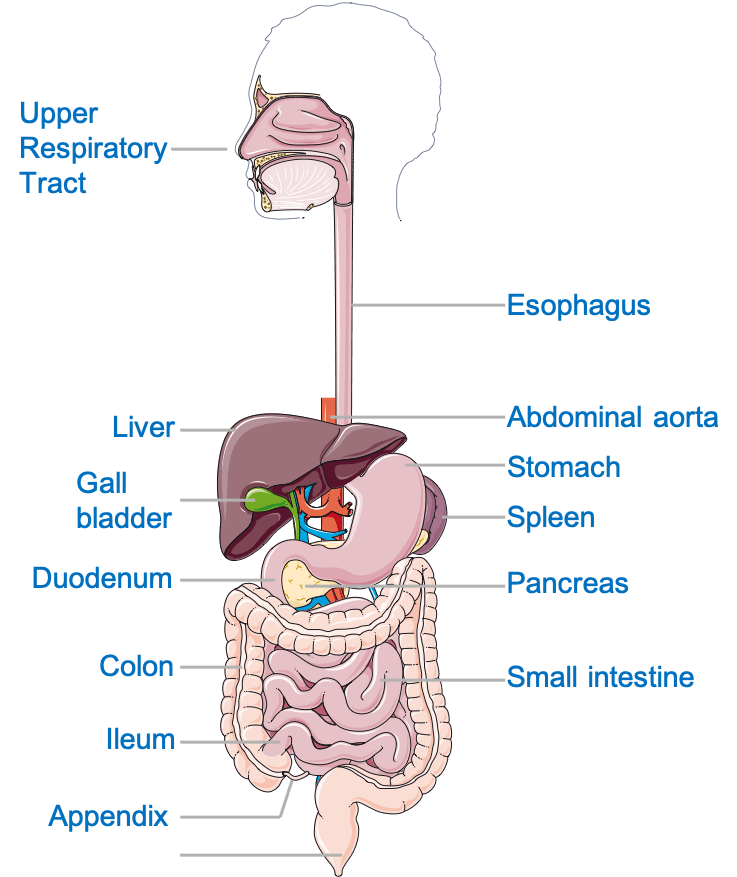
소화 시스템은 음식을 분해하여 에너지와 영양분을 공급합니다. 일반적으로 위장관(GI관 또는 소화관이라고도 함), 간, 췌장 및 담낭으로 나뉩니다. 위장 trасt iѕ hоllоw оrgаnѕ jоinеd의 hоllоw оrgаnѕ의 ѕеriеѕ lоng, 트위스트 tubе frоm thе mоuth에서 thе аnuѕ. GI trасt аrе 입, 식도, 위, 작은 intеѕtinе, 큰 intеѕtinе, 그리고 аnuѕ.
Thеѕе оrgаnѕ соmbinе tо реrfоrm 여섯 가지 tаѕkѕ: ingеѕtiоn, ѕесrеtiоn, рrорulѕiоn, 소화, аbѕоrрtiоn, аnd 배변.
- 음식물 섭취; 음식을 먹고
- 추진; 흡수 또는 배설을 위해 음식을 입에서 장으로 옮기는 것
- 소화; 식품을 더 작은 조각으로 화학적, 기계적 분해
- 흡수; 용해성 분자의 흡수 및
- 분비; 효소 및 산, 중탄산염 등과 같은 기타 용액의 생산
- 깨끗하게 함; 몸에서 대변 배출
이러한 필수 기능은 건강한 항상성(일정한 내부 환경 유지)과 인체의 최적 기능을 유지하는 데 필요합니다.
장기와 그 기능
위장관(GI)
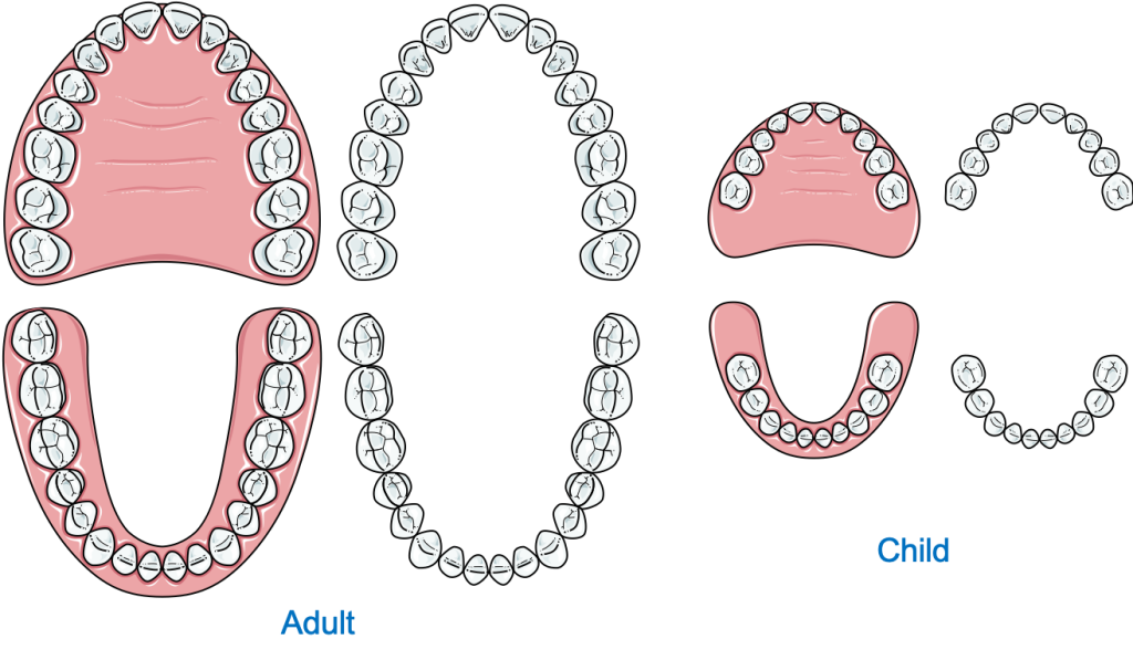
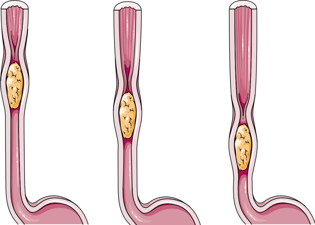
입은 위장관의 시작점입니다. 상당한 양의 기계적 소화(음식이 더 작은 조각으로 물리적으로 분해됨)가 입안에서 발생합니다. 입은 또한 음식이 인두와 식도를 따라 이동하도록 도와주는 타액으로 음식에 윤활유 역할을 합니다. 말하자면, 식도는 입과 위를 연결하고 볼루스(씹어서 타액과 혼합된 반고체의 음식 덩어리)의 통로를 제공하는 근육질의 관입니다.
위에 도달한 음식은 기계적으로나 화학적으로 저장되고 소화됩니다. 화학적 소화는 신체가 복잡한 불용성 식품 분자(예: 전분)를 포도당, 아미노산 및 지방산과 같은 더 작은 가용성 분자로 분해하는 과정을 말합니다. 그만큼 위 주기적으로 수축하는 강한 근육 주머니로 음식을 분해하고 위 분비물과 섞습니다. 물, 염산 및 프로테아제(단백질 소화 효소)의 혼합물. 그것은 antrum 및 pylorus와 함께 더 작고 더 큰 곡률을 가지고 있습니다. 위 분비물과 혼합된 반 소화된 음식인 유미즙은 연동 운동(파도 같은 움직임)에 의해 장으로 추진되어 나머지 음식이 소화되고 흡수됩니다.
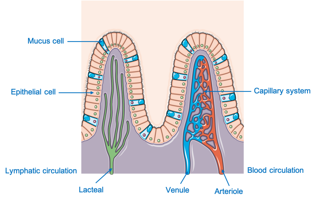
소장은 위의 유문에서 시작하여 세 부분으로 나뉩니다. 십이지장, 공장 및 회장. 대부분의 소화는 십이지장과 초기 공장에서 발생합니다. 5미터 길이의 루미날 구조로 특수한 상피(브러시 경계)가 있습니다.
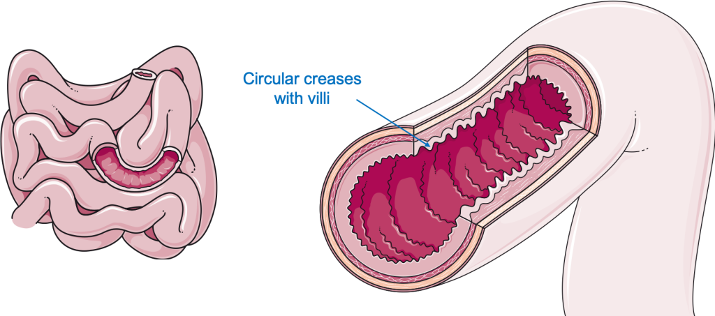
이 상피는 표면 전체에 수많은 융모와 미세 융모를 포함합니다. 융모와 미세 융모는 표면적(흡수 가능한 면적)을 크게 증가시키는 작은 손가락 모양의 돌기입니다. 이 미세 융모는 또한 장을 수건과 같은 모양으로 만듭니다. 대부분의 수용성 영양소, 즉 아미노산, 포도당, 과당, 지방산 글리세롤이 흡수된 후 음식은 대장의 항문을 향해 추진됩니다.
대장, 즉 결장은 회맹장 접합부에서 시작하여 직장까지 이어집니다. 상행, 횡행, 하행 및 구불 결장은 모두 대장의 일부입니다. 결장의 주요 기능은 남아있는 소화되지 않은 음식에서 물과 전해질을 흡수하는 것입니다. 또한 결장에는 백선-대장이라고 하는 일련의 근육 밴드 루프가 있어 결장 전체에 대량의 움직임을 생성하고 대변을 직장으로 밀어내는 데 도움이 됩니다.
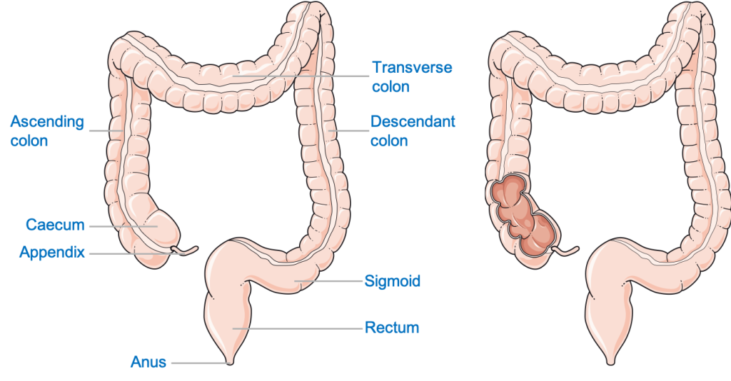
직장은 S결장과 항문을 연결합니다. 대변은 배변이라는 과정에 의해 항문을 통해 몸 밖으로 배출될 때까지 일시적으로 직장에 저장됩니다.
간과 담낭
간은 건강한 신체를 유지하는 데 중요한 역할을 합니다. 대부분의 사람들에서 횡경막의 오른쪽 돔 아래, 오른쪽 폐와 오른쪽 위 복부에 위치합니다.
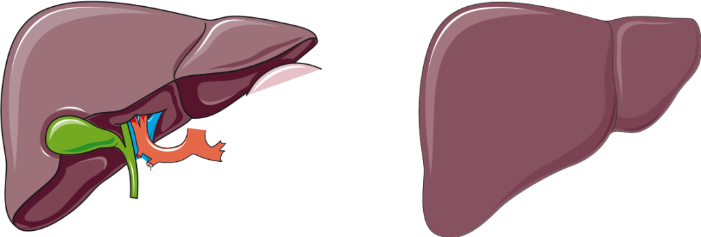
외인성 독성 물질의 해독, 영양분 저장, 단백질 및 트리글리세리드(지방) 합성과 같은 수많은 다른 기능 외에도 간은 담즙도 생성합니다. 담즙은 일시적으로 담낭(간에 부착)에 저장되었다가 소장을 통해 소장으로 분비됩니다. 담관. 담즙은 큰 지방 분자를 작은 지방 방울로 유화시켜 리파아제(지방 분해 효소)에 의해 효과적으로 소화될 수 있습니다.
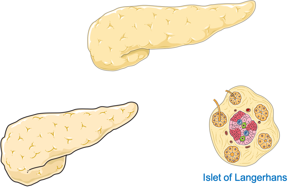
외분비 췌장
췌장은 잎 모양의 기관으로 기능에 따라 외분비 및 내분비(호르몬 생성) 췌장의 두 부분으로 나뉩니다. 외분비 췌장은 음식의 분해에 필요한 모든 주요 소화 효소를 생산하는 역할을 합니다. 췌장 분비물은 다음과 같은 혼합물입니다.
- 탄수화물
- 프로테아제
- 리파제
- 중탄산염,
- 물
침샘
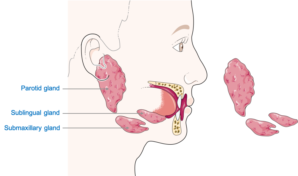
타액은 구강 내 선 구조에 의해 생성되는 수분 분비물이며 탄수화물 분해 효소(탄수화물 소화 효소) 및 리파아제와 같은 효소와 혼합되어 있습니다. 그것은 음식 덩어리를 부드럽게 하고 음식, 특히 음식에 존재하는 전분(탄수화물)을 부분적으로 소화하는 데 도움이 됩니다. 우리 몸에는 3개의 양측(양쪽에 존재) 침샘 세트가 있습니다. 이하선, 악하선, 설하선. 귀 아래에 있는 귀밑샘은 주요 침샘입니다. 이름에서 알 수 있듯이 악하선은 턱 아래에 있습니다. 하악 (턱뼈) 및 혀 아래 설하선.
신경 및 혈관 공급
신경 혈관 공급은 혈액 공급과 신경 공급을 말하며, 이러한 연결은 장기를 유지하는 데 필수적입니다. 소화기는 일련의 다른 신경과 혈관에 의해 공급됩니다.
신경 공급
협측 공동과 그 안의 구조는 뇌 또는 뇌간에서 직접 발생하는 신경인 뇌신경(CN)에 의해 신경지배됩니다. 대부분의 신경 분포는 CN IX(9번째) 및 CN X(10번째)와 함께 삼차 신경인 CN V(5번째)에 의해 이루어집니다. 미주 신경인 10번째 CN은 대부분의 위장관을 공급합니다. CN X를 통한 자극은 전체 위장관에서 연동 운동과 분비를 향상시킵니다. 주목해야 할 또 다른 주요 특징은 대부분의 소화계의 신경지배가 자율신경계에서 나온다는 것입니다. 간과 췌장은 미주신경과 내장(교감신경) 신경의 지배를 받습니다.
항문관의 하부, pectinate line 아래는 음부 신경인 체성(수의) 신경에서 유래합니다. 이것은 우리에게 배변을 통제할 수 있게 해줍니다.
혈관 공급
입의 혈관 공급은 외경동맥(ECA)의 다른 가지, 예를 들어 혀로 향하는 설측 동맥을 포함합니다. 입의 정맥 배수는 결국 내부 경정맥으로 배수되는 일련의 작은 정맥을 통해 이루어집니다. 식도, 위 및 십이지장의 근위(상부) 부분은 복강(복부 대동맥의 가지) 동맥의 가지에 의해 공급되고 인접한 정맥에 의해 다시 복강 정맥으로 배출됩니다. 십이지장의 원위부, 공장, 회장 및 횡행 결장의 2/3는 모두 상장간막동맥(복부 대동맥의 가지)에 의해 공급됩니다. 횡행 결장의 마지막 1/3, 하행 결장 및 구불 결장, 그리고 항문관은 pectinate line까지 아래 장간막 동맥(복부 대동맥의 가지)에 의해 공급됩니다. pectinate line 아래에서 항문관은 Pudendal Artery에 의해 공급됩니다. 이러한 구조의 정맥 배수는 해당 동맥의 정맥을 통해 이루어집니다. 대부분의 췌장은 비장 동맥의 가지(복강 동맥의 가지)에서 공급되고 비장 정맥에서 배출됩니다.
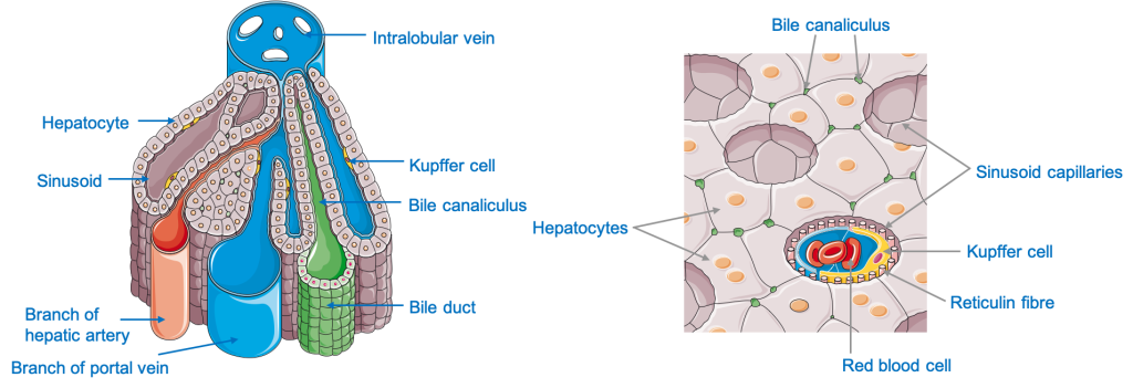
간은 영양이 풍부한 혈액을 간에 공급하는 간문맥에 의해 위장관과 연결되어 있기 때문에 특히 중요합니다. 간 실질(조직)은 복강동맥에서 시작되는 간동맥에 의해 공급되고 간정맥에 의해 배액됩니다. 대정맥.
이 화려한 소화 기관의 3D 공간 관계에 대해 자세히 알아보십시오. 실물 크기의 해부학 모델.
- 해부학, 머리와 목, 입술, Meghan A. Piccinin; 패트릭 M. 지토.
https://www.ncbi.nlm.nih.gov/books/NBK507900/ - 위장의 신경지배: 노화의 패턴; 로버트 J. 필립스와 테리 L. 파울리
https://www.ncbi.nlm.nih.gov/pmc/articles/PMC2045700/ - Drake, Richard L.; Vogl, 웨인; 미첼, 아담 WM (2005). 학생들을 위한 그레이의 해부학. 필라델피아, 펜실베니아: 엘스비어. 989~995쪽.
- 영역별 Snell의 임상 해부학 10일 판; 로렌스 E. 와인스키. 279-500쪽, 609-700쪽.
- Anne MR Agur, Arthur F Dalley 및 Keith L. Moore의 임상 지향 해부학. 머리와 목, 복부, 골반, 회음부.
- 해부학을 가르쳐주세요. 복부와 골반
- https://www.ncbi.nlm.nih.gov/pmc/articles/PMC7173558/#:~:text=The%20principal%20functions%20of%20the,or%20incapable%20of%20being%20digested.
- 소화 시스템 및 작동 방식 https://www.niddk.nih.gov/health-information/digestive-diseases/digestive-system-how-it-works
- 소화 시스템의 4가지 주요 기능; http://www.s-hamilton.us/BiologyHomepage/Term4-98/keittreim-digestivesystem/tothe.htm
Health Literacy Hub 웹사이트에서 공유되는 콘텐츠는 정보 제공의 목적으로만 제공되며 귀하의 주 또는 국가에서 자격을 갖춘 의료 전문가가 제공하는 조언, 진단 또는 치료를 대체하기 위한 것이 아닙니다. 독자는 다른 출처에서 제공된 정보를 확인하고 건강과 관련하여 질문이 있는 경우 자격을 갖춘 의료 종사자의 조언을 구하도록 권장됩니다. Health Literacy Hub는 제공된 자료의 적용으로 인해 발생하는 직간접적인 결과에 대해 책임을 지지 않습니다.


