3D Anatomy Model
Add another dimension to your learning with fully-interactive educational male and female anatomical models.
Learning about the human anatomy has never been more fun!
Purchase
Corpus Callosum is fundamentally a bundle of different types of nerve fibers in the brain.
Let’s learn some interesting facts about the corpus callosum:
1. Corpus callosum derives from Latin and means “tough body”, due to its nature of tight nerve fibers bonded together.
2. The corpus callosum of a person contains approximately more than 200 million axons, making it the largest connective pathway in the brain.
3. This bundle of fibers connects the right and left brain hemispheres. The corpus callosum ensures that the right and left cerebral hemispheres communicate with each other.
4. The nerve fibers of the corpus callosum are of different types. Hence, there is the transfer of all kinds of signals, i.e., sensory, motor, and cognitive signals, between two hemispheres of the human brain through the corpus callosum.
5. Brain abnormalities that interest the corpus callosum can occur during fetal brain development due to chromosomal abnormality, environmental or viral causes. Callosal agenesis and callosal dysgenesis refer to the partial or complete absence, or abnormal development, of the corpus callosum. Corpus callosum malformation manifests with developmental delay and cognitive impairment.
The following section includes a detailed description of the corpus callosum’s structure, functions, blood supply, and clinical conditions.
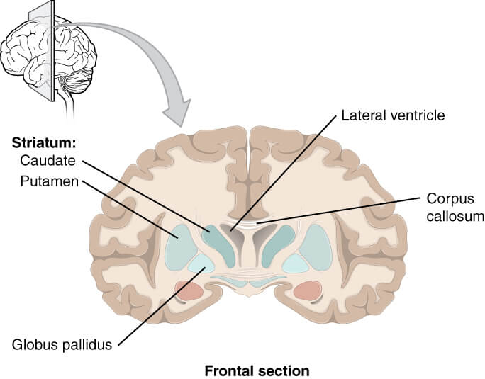
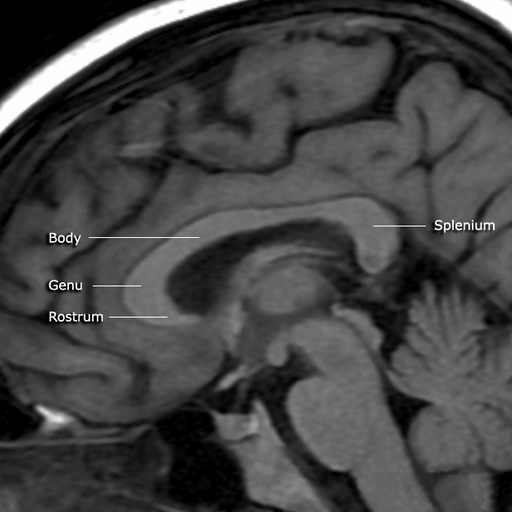
Carpus Callosum is a large band of white matter. It is the largest white matter structure of the whole brain, both in size and number of neurons. The approximate size of the corpus callosum is 700 square millimetres and contains more than 200 million axons. Carpus Callosum is divided into four parts. These parts include the rostrum, genu, and splenium of the carpus Callosum from the anterior to the posterior sequence.

The rostrum is the anteroinferior part of the carpus callosum. The rostrum of the carpus callosum is continuous with the lamina terminals. The lamina terminals connect the corpus callosum with the frontal lobe of the brain at the orbital surface.
The genu is the anterior bent part of the corpus callosum. The fibers projecting from the genu of the corpus callosum connect different parts of the frontal lobe.
The body or trunk of the corpus callosum starts from the genu and ends at the splenium. The fibers original from this central section of the carpus callosum traverse through the corona radiate to approach the surface of the right and left hemispheres.
The splenium is the posterior tapered part of the corpus callosum. The fibers original from the splenium is destined to connect the right and left occipital lobe.
The primary function of the corpus callosum is to create communication between the right and left hemispheres. The corpus callosum connects the similar areas of two hemispheres, which allows those parts to communicate and work in harmonization. Different parts of the carpus callosum create communication between the areas of the hemispheres.
Besides the primary function of the corpus callosum, it is hypothesized that it plays a significant role in cognition. Recent researches indicate that corpus callosum has a role in intelligence, thinking, speed, and problem-solving instincts. However, there is a need for more research to create a clear relationship between the corpus callosum and the process of cognition.
Corpus Callosum is supplied by several arteries that anastomose with each other. Hence it has a rich blood supply and is usually safe from infarction. Following vessels have contributed to the blood supply of the corpus callosum.
The pericallosal artery runs along the outer border of the corpus callosum, initially in the subcallosal area and then through the callosal or cingulate sulcus. The branches from this artery supply blood to most of the corpus callosum.
The splenial or posterior pericallosal artery usually arises from the posterior cerebral artery. It divides into superior and inferior branches that supply blood to the respective aspects of the splenium of the corpus callosum. Branches from the splenial artery are usually anastomose with the branches of the pericallosal artery.
In most people, the anterior communicating artery supplies blood to the corpus callosum through the subcallosal or median callosal artery. The subcallosal artery supplies blood to the hypothalamus along with the rostrum and genu of the corpus callosum. The median callosal artery also follows the same route, but it extends up to the body of the corpus callosum.
The venous drainage of the corpus callosum occurs through the callosal and callosocingulate veins. The two veins join at the central level of the corpus callosum to form subependymal veins which drain into the septal veins and median atrial veins. All these veins ultimately drain into internal cerebral veins.
In broad terms, there are two types of connections created by the corpus callosum. The homotopic connections that link the same parts of the brain from the right and left side. And the heterotopic connections that connect different parts of the brain from the right and left sides. Various terms are used to describe the fibers originating from different parts of the corpus callosum.
Forceps minor is the tract that arises from the genu of the corpus callosum and connects the parts of the frontal lobe among them.
Forceps major is the tract that arises from the splenium of the corpus callosum and connects the right and left occipital lobes.
Tapetum is the large sheet of white matter that projects from the body of the corpus callosum and connects the right and left hemispheres. The fibers arising from the splenium that do not connect the right and left occipital lobe are also part of the tapetum.
Agenesis of the carpus Callosum is a rare congenital disorder in which the corpus callosum is entirely absent. It occurs between the 3rd and 12th week of gestation, the period of peak neurological development.
The patients of agenesis of corpus callosum present with a complex clinical picture. The symptoms include poor motor coordination, hypotonia, difficulty swallowing, poor pain perception, and cognitive impairment. There are also seizures in some cases of agenesis of the corpus callosum. The specific symptoms of the disease vary according to the co-existing neurological conditions.
A long-standing hydrocephalus cause impingement of the white matter of the corpus callosum on the free inferior margin of the falx cerebri. This impingement of the corpus callosum can lead to ischemia and atrophy. Corpus Callosum Impingement syndrome is usually asymptomatic in most cases.
The Corpus Callosum Disconnection Syndrome is characterized by a disconnection between the right and left hemispheres due to stroke, surgery, or trauma. The patient might have normal routine performance, but the symptoms of abnormality appear when there is increased pressure on the brain during specific tests. The patients with disconnection syndrome have difficulty in an organized speech during the specific tests.
Corpus callosum is a large band of white matter that lies deep to the cingulate gyrus. The primary function of the corpus callosum is to create communication between the right and left sides of the brain. It also plays some role in cognition. There are four parts of the corpus callosum; rostrum, genu, body, and splenium.
The blood supply of the corpus callosum is from the anterior communicating artery, pericallosal artery, and posterior pericallosal artery. These arteries anastomose among them, which reduces the risk of ischemia in the corpus callosum.
The clinical conditions related to corpus callosum include its agenesis and impingement. The symptoms of corpus callosum disorders include speech disruption, poor motor coordination, difficulty swallowing, etc. The specific symptoms of the disease depend on the co-existing neurological conditions.
1. Goldstein, A., Covington, B. P., Mahabadi, N., & Mesfin, F. B. (2020). Neuroanatomy, Corpus Callosum. In StatPearls. StatPearls Publishing. https://pubmed.ncbi.nlm.nih.gov/28846239/
2. Roland, J. L., Snyder, A. Z., Hacker, C. D., Mitra, A., Shimony, J. S., Limbrick, D. D., Raichle, M. E., Smyth, M. D., & Leuthardt, E. C. (2017). On the role of the corpus callosum in interhemispheric functional connectivity in humans. Proceedings of the National Academy of Sciences of the United States of America, 114(50), 13278–13283. https://doi.org/10.1073/pnas.1707050114
3. Goldstein A, Covington BP, Mahabadi N, et al. Neuroanatomy, Corpus Callosum. [Updated 2020 Jul 31]. In: StatPearls [Internet]. Treasure Island (FL): StatPearls Publishing; 2021 Jan-. Available from: https://www.ncbi.nlm.nih.gov/books/NBK448209/
4. Hofman, J., Hutny, M., Sztuba, K., & Paprocka, J. (2020). Corpus Callosum Agenesis: An Insight into the Etiology and Spectrum of Symptoms. Brain sciences, 10(9), 625. https://doi.org/10.3390/brainsci10090625
5. Murthy, S. B., Chmayssani, M., Shah, S., Goldsmith, C. E., & Kass, J. S. (2013). Clinical and radiologic spectrum of corpus callosum infarctions: clues to the etiology. Journal of clinical neuroscience : official journal of the Neurosurgical Society of Australasia, 20(1), 175–177. https://doi.org/10.1016/j.jocn.2012.05.013
The content shared in the Health Literacy Hub website is provided for informational purposes only and it is not intended to replace advice, diagnosis, or treatment offered by qualified medical professionals in your State or Country. Readers are encouraged to confirm the information provided with other sources, and to seek the advice of a qualified medical practitioner with any question they may have regarding their health. The Health Literacy Hub is not liable for any direct or indirect consequence arising from the application of the material provided.


