3D Anatomy Model
Add another dimension to your learning with fully-interactive educational male and female anatomical models.
Learning about the human anatomy has never been more fun!
Purchase
The brain is the most vital organ of the body, and the function of the whole body depends on the functioning of the brain. As per the importance of the brain, it needs to be more protected than the other organs of our body. The outermost protection of the brain is a skull. For further protection of the brain, there are three layers of connective tissue beneath the skull that protect the brain from injury and keep it at the position even if there is a limited skull injury.
The brain’s protective connective tissue layers are meninges, including the pia mater, arachnoid mater, and dura mater. The pia mater is the innermost, thin, and delicate layer attached to the brain and follows the contour and involutions of the brain. The arachnoid mater is outer to the pia mater and does not follow the curves and shape of the brain.
Some interesting facts about the dura mater include:
1. The cranial dura is the outermost and toughest layer of the meninges and has a highly protective function for the brain.
2. The spinal cord is also part of the central nervous system and is protected similarly to the brain. However, instead of the skull, the spinal cord has the protection of the vertebral column. The space between the arachnoid mater and the pia matermeninges is called subarachnoid space, and it contains the cerebrospinal fluid (CSF). In the case of dural tear, or damage of the spinal dura, the CSF can leak out of this space.
3. Amongst the three layers of meninges, the Dura mater has the largest blood vessels.
In this article we will discuss the structure and function of the cranial and spinal dura, and the complications arising from injury to this meningeal layer.
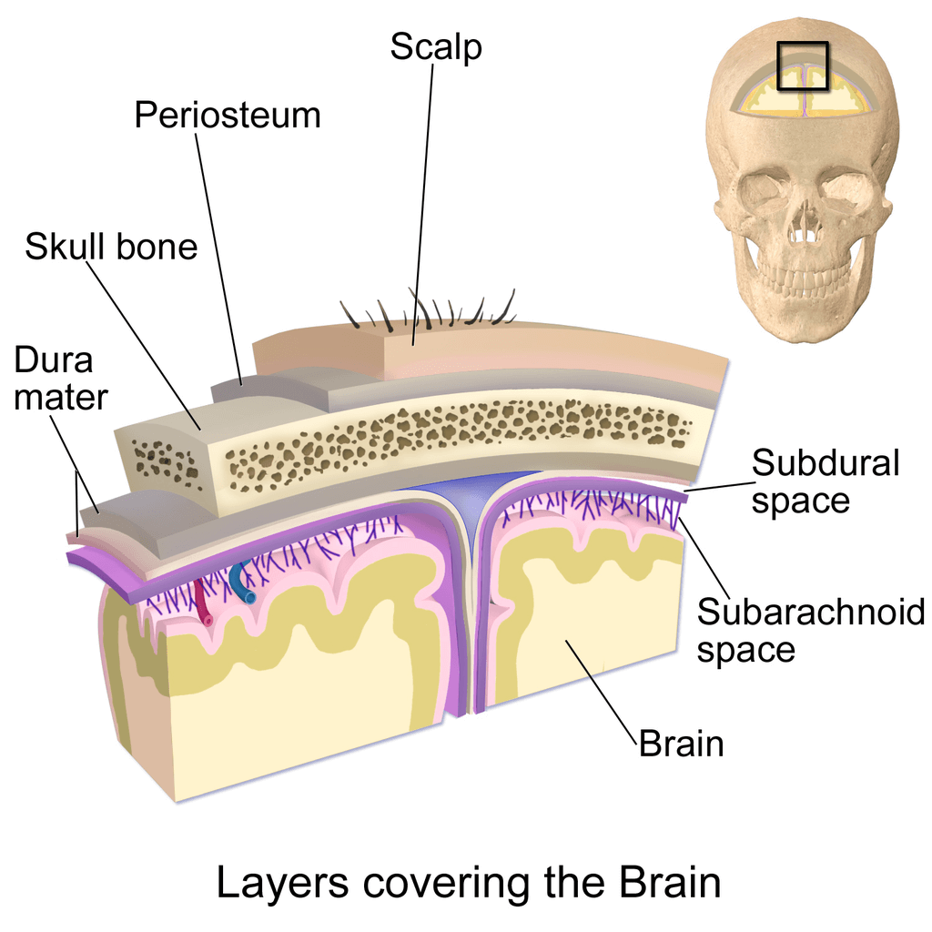
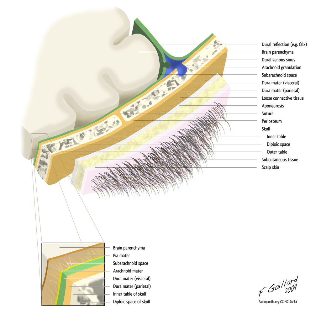
The Dura is the outermost layer of the meninges that covers the brain and the entire length of the spinal cord. The Dura mater is located immediately beneath the skull and bones of the vertebral column. Dura is thick in structure which makes it tough and inextricable.
The Dural layer of meninges itself consists of two further layers of connective tissue.
The periosteal layer is the outer layer of durra that is like the inner lining of the skull.
A meningeal layer of Dura lies deeper into the periosteal layer. The meningeal layer of the dura mater of the brain and spinal cord is continuous.
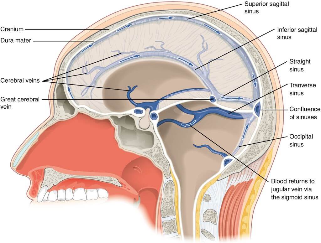
The space between the two layers of Dura has room for the venous drainage of the cranium. These spaces are termed dural venous sinuses. The dural venous sinuses drain the venous blood from the cranium and open it into the internal jugular veins.
Four dural reflections are formed by the inward folding of the meningeal layer of Dura around itself. The dural reflections project in the cranial cavity is creating different compartments of the cranial cavity. Different parts of the brain occupy the compartments or divisions of the cranial cavity.
There are four major reflections of the Dura.
Falx cerebri projects from the middle of the Dura and separates the right and left cerebral hemispheres.
The tentorium cerebelli is the dural reflection that separates the cerebellum and the occipital lobe. In the anteromedial part of the tentorium, there is space for the passage of the midbrain through it. This space or notch of the tentorium is called a tentorial notch.
Falx cerebelli acts as a separation between the right and left cerebellar hemispheres.
Diaphragm Sellae is another dural projection that acts as a tent-like covering over the hypophysial fossa of the sphenoid bone. The stalk of the pituitary gland passes through a small opening of the diaphragm sellae.
The dura mater is a tough membrane and protects the brain from traumatic injuries. Whenever there is some accident or blow on the scalp, the Dura keeps the brain at its place and does not let it reach the hard bony structure of the brain. If the Dura and other layers of meninges do not support the brain, it will hit the hard scalp whenever there is external trauma. This hitting of the brain on the hard scalp will result in traumatic injuries.
The Dura and other layers of meninges act as a pathway through which the blood vessels traverse to read and supply blood to the brain. The dural venous sinuses also help in draining the venous blood from the brain and cranium.
There are also some other functions of Dura which include:
The Dura of different brain and spinal cord parts has blood supply from branches of different blood vessels. The vessels that are the main source of blood for the dura mater include the internal carotid artery, maxillary artery, occipital artery, ascending pharyngeal artery, and branches of the vertebral artery.
The venous drainage of the Dura occurs through the meningeal veins. Meningeal veins are present in the periosteal layer. The dura mater and follow the course corresponding branches of the middle meningeal artery. The meningeal veins ultimately drain into the sphenopalatine sinus. They may also drain through the pterygoid venous plexus.
The neural innervation of the dura mater is through branches of the sympathetic nervous system:
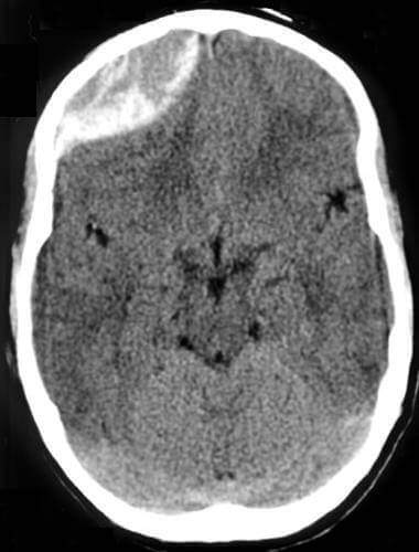
The common clinical condition related to Dura is hemorrhage formation of a hematoma. Hematoma can be described as a bad bruise or collection of blood. As the cranial cavity is a closed space with limited capacity, hematoma formation leads to raised intracranial pressure and subsequent damage. The hemorrhage and hematoma formation related to the dura mater is divided into two types.
The hemorrhage or hematoma formation between the bones of the skull and periosteal layer of the dura mater is called extradural hematoma/hemorrhage. The extradural hemorrhage and hematoma formation occur due to rupture of the middle meningeal artery.
A subdural hematoma formation occurs due to rupture of the cerebral veins that empty into dural sinuses. In subdural hematoma, there is an accumulation of blood between the dura mater and arachnoid mater.
Dura is the outermost and toughest layer of the meninges. It consists of two connective tissue layers, the periosteal and meningeal layers. Four dural projections arise from the Dura that separates the different parts of your brain.
The main function of the Dura is to protect the brain against traumatic injury. However, there are some other functions, such as providing a framework for the blood vessels. Various blood vessels supply the blood to the Dura. The venous drainage of the Dura occurs through meningeal veins into the sphenopalatine sinus. The nerve supply of the Dura is also from a mixture of cranial and spinal nerves.
Two common clinical conditions related to Dura include extradural and subdural hematoma. The results of these hematomas are severe due to brain damage.
1: Kekere, V., & Alsayouri, K. (2020). Anatomy, Head and Neck, Dura Mater. In StatPearls. StatPearls Publishing. https://pubmed.ncbi.nlm.nih.gov/31424885/
2: Kekere V, Alsayouri K. Anatomy, Head and Neck, Dura Mater. [Updated 2020 Aug 10]. In: StatPearls [Internet]. Treasure Island (FL): StatPearls Publishing; 2021 Jan-. Available from: https://www.ncbi.nlm.nih.gov/books/NBK545301/
3: Zwirner J, Scholze M, Waddell JN, Ondruschka B, Hammer N. Mechanical Properties of Human Dura Mater in Tension – An Analysis at an Age Range of 2 to 94 Years. Sci Rep. 2019;9(1):16655. Published 2019 Nov 13. doi:10.1038/s41598-019-52836-9 https://www.ncbi.nlm.nih.gov/pmc/articles/PMC6853942/
4: Lv, X., Wu, Z., & Li, Y. (2014). Innervation of the cerebral dura mater. The neuroradiology journal, 27(3), 293–298. https://doi.org/10.15274/NRJ-2014-10052
5: Weller, R. O., Sharp, M. M., Christodoulides, M., Carare, R. O., & Møllgård, K. (2018). The meninges as barriers and facilitators for the movement of fluid, cells and pathogens related to the rodent and human CNS. Acta neuropathologica, 135(3), 363–385. https://doi.org/10.1007/s00401-018-1809-z
6: Mack, J., Squier, W., & Eastman, J. T. (2009). Anatomy and development of the meninges: implications for subdural collections and CSF circulation. Pediatric radiology, 39(3), 200–210. https://doi.org/10.1007/s00247-008-1084-6
The content shared in the Health Literacy Hub website is provided for informational purposes only and it is not intended to replace advice, diagnosis, or treatment offered by qualified medical professionals in your State or Country. Readers are encouraged to confirm the information provided with other sources, and to seek the advice of a qualified medical practitioner with any question they may have regarding their health. The Health Literacy Hub is not liable for any direct or indirect consequence arising from the application of the material provided.


